| Issue |
Parasite
Volume 32, 2025
|
|
|---|---|---|
| Article Number | 37 | |
| Number of page(s) | 14 | |
| DOI | https://doi.org/10.1051/parasite/2025030 | |
| Published online | 24 June 2025 | |
Research Article
A one-step multiplex qPCR assay for simultaneous identification and quantification of Leishmania martiniquensis and Leishmania orientalis/Leishmania chancei and detection and quantification of trypanosomatids in clinical samples
Un test de qPCR multiplex en une étape pour l’identification et la quantification simultanées de Leishmania martiniquensis et Leishmania orientalis/Leishmania chancei, ainsi que pour la détection et la quantification des trypanosomatidés dans les échantillons cliniques
1
Center of Excellence in Vector Biology and Vector-Borne Disease, Department of Parasitology, Faculty of Medicine, Chulalongkorn University, Bangkok 10330, Thailand
2
Department of Medical and Public Health Secretary, College of Allied Health Sciences, Suan Sunandha Rajabhat University, Samut Songkhram 75000, Thailand
3
Faculty of Medicine, Chulalongkorn University, Bangkok 10330, Thailand
4
Department of Internal Medicine, Faculty of Medicine, Chiang Mai University, Chiang Mai 50200, Thailand
5
Division of Biomedical and Life Sciences, Faculty of Health and Medicine, Lancaster University, Lancaster LA1 4YG, UK
* Corresponding author: This email address is being protected from spambots. You need JavaScript enabled to view it.
Received:
5
December
2024
Accepted:
26
May
2025
Abstract
Leishmaniasis is one of the most important zoonotic diseases. Recently, Leishmania (Mundinia) martiniquensis, Leishmania (Mundinia) orientalis, and Leishmania (Mundinia) chancei have been reported as new human pathogens. Trypanosomatids, apart from Leishmania spp., such as Crithidia spp., are also occasionally capable of infecting humans. Here, a one-step multiplex qPCR assay for the simultaneous identification and quantification of L. martiniquensis and L. orientalis/L. chancei and detection and quantification of trypanosomatids was developed using ITS1 as the molecular target and human RNase P as the internal control gene. The assay was evaluated using 44 positive residual DNA samples from leishmaniasis patients and 25 negative DNA samples. Results revealed that the limits of detection (LOD) of the assay for L. martiniquensis, L. orientalis, and Crithidia sp. (CLA-KP1) were 1.699 (0.0255), 1.717 (0.0292), and 1.763 (0.0882) fg/reaction (parasite equivalents/reaction), respectively. The assay had high analytical specificity. The mean Cq values of the intra-assays and inter-assays differed by less than 1, indicating the reliability of the assay. Evaluation results revealed that the assay could identify L. martiniquensis and L. orientalis in clinical samples with 100% sensitivity and 100% specificity. In conclusion, the ITS1/human RNase P multiplex qPCR assay offers a rapid and reliable diagnostic tool for identifying and quantifying L. martiniquensis and L. orientalis/L. chancei and detecting and quantifying trypanosomatids in clinical samples within a single reaction. This assay provides an advancement in the diagnostic capabilities for leishmaniasis and trypanosomatid infections, potentially improving patient management and surveillance efforts.
Résumé
La leishmaniose est l’une des zoonoses les plus importantes. Récemment, Leishmania (Mundinia) martiniquensis, Leishmania (Mundinia) orientalis et Leishmania (Mundinia) chancei ont été signalés comme de nouveaux agents pathogènes humains. Outre Leishmania spp., les trypanosomatidés, tels que Crithidia spp., sont également parfois capables d’infecter l’homme. Un test de qPCR multiplex en une étape pour l’identification et la quantification simultanées de L. martiniquensis et de L. orientalis/L. chancei, ainsi que pour la détection et la quantification des trypanosomatidés, a été développé en utilisant ITS1 comme cible moléculaire et la RNAase P humaine comme gène de contrôle interne. Le test a été évalué à l’aide de 44 échantillons d’ADN résiduel positifs provenant de patients atteints de leishmaniose et de 25 échantillons d’ADN négatifs. Les résultats ont révélé que les limites de détection (LOD) du test pour L. martiniquensis, L. orientalis et Crithidia sp. (CLA-KP1) étaient respectivement de 1,699 (0,0255), 1,717 (0,0292) et 1,763 (0,0882) fg/réaction (équivalents parasites/réaction). Le test présentait une spécificité analytique élevée. Les valeurs moyennes de Cq des tests intra-tests et inter-tests différaient de moins de 1, ce qui indique la fiabilité du test. Les résultats de l’évaluation ont révélé que le test pouvait identifier L. martiniquensis et L. orientalis dans des échantillons cliniques avec une sensibilité et une spécificité de 100 %. En conclusion, le test qPCR multiplex ITS1/RNAase P humaine offre un outil de diagnostic rapide et fiable pour l’identification et la quantification de L. martiniquensis et L. orientalis/L. chancei, ainsi que pour la détection et la quantification des trypanosomatidés dans les échantillons cliniques, en une seule réaction. Ce test constitue une avancée dans le diagnostic de la leishmaniose et des infections par les trypanosomatidés, améliorant potentiellement la prise en charge des patients et la surveillance.
Key words: qPCR / Leishmania martiniquensis / Leishmania orientalis / Leishmania chancei / Mundinia / trypanosomatid
Edited by: Jérôme Depaquit
© C. Mano et al., published by EDP Sciences, 2025
 This is an Open Access article distributed under the terms of the Creative Commons Attribution License (https://creativecommons.org/licenses/by/4.0), which permits unrestricted use, distribution, and reproduction in any medium, provided the original work is properly cited.
Introduction
Leishmaniasis is an important vector-borne zoonotic disease that affects an estimated 350 million individuals living in tropical and subtropical areas [72]. It is caused by protozoan parasites of the genus Leishmania, which belong to the Trypanosomatidae family and can infect humans as well as several domestic and sylvatic animals [6, 10, 29, 37, 42, 44, 57]. According to the World Health Organization, an estimated 700,000 to 1 million new cases of leishmaniasis occur annually, leading to significant morbidity and mortality, especially in underdeveloped countries [72]. The clinical manifestations of leishmaniasis vary depending on the species and/or strain of Leishmania and host immune response. Disease severity ranges from asymptomatic or self-healing cutaneous lesions (cutaneous leishmaniasis, CL) to chronic infections that cause severe tissue destruction in mucocutaneous leishmaniasis (MCL) and can progress to life-threatening visceral leishmaniasis (VL) [72]. Various species of sand flies are well-established vectors of Leishmania parasites in the subgenera Leishmania and Viannia. However, recent reports have documented natural infection of the human pathogen Leishmania (Mundinia) martiniquensis in Culicoides peregrinus, suggesting that biting midges may serve as potential vectors of leishmaniasis [22]. Moreover, transmission through blood transfusion may also be possible, as asymptomatic Leishmania carriers have been identified among blood donors in several endemic countries, including Brazil and Iran [4, 64]. Recently, at least 21 Leishmania species have been identified as human pathogens, primarily belonging to the subgenera Leishmania (ten species), Viannia (eight species), and the newly identified subgenus Mundinia (three species) [62]. The emergence of novel human infective Leishmania species of subgenus Mundinia, including L. (Mundinia) martiniquensis [8], L. (Mundinia) orientalis [20], and L. (Mundinia) chancei [25], poses an increasing public health and disease control challenge in endemic areas.
Thailand is considered endemic for leishmaniasis, particularly in the northern and southern parts of the country, with confirmed autochthonous cases and a continuously rising number of both symptomatic and asymptomatic cases over the years [5, 27, 53, 63, 66, 72]. This disease is caused by two main protozoan species including L. martiniquensis [52] and L. orientalis [20]. In an immunocompetent host, L. martiniquensis causes VL [52], while L. orientalis is mainly responsible for CL [20]. However, VL, diffuse CL, and MCL have been observed in immunocompromised hosts infected with L. martiniquensis [66]. Other Leishmania species documented in Thailand include L. donovani complex, L. lainsoni, L. major, and L. infantum [30, 33]. Recently, amphotericin B-resistant L. martiniquensis parasites have also been reported in Thailand [21, 31, 32]. For another L. (Mundinia) species, L. chancei (formerly L. “Ghana”), which belongs to a new subgenus and is endemic to West Africa, also causes CL characterized by skin lesions, often ulcers, that can be accompanied by satellite lesions or nodular lymphangitis [26]. The varied clinical manifestations of leishmaniasis caused by L. martiniquensis, L. orientalis, and L. chancei require specific therapeutic and treatment strategies for each species. Furthermore, most of these infection cases are asymptomatic Leishmania/HIV co-infections, which pose a major challenge to diagnosing and controlling the disease [33, 67]. The absence of clinical lesions in these individuals, especially in VL/HIV cases, increases the risk of treatment failure/drug resistance and may contribute to the transmission cycle and spread of the disease [2, 39]. International travel and migration by asymptomatic individuals to and from endemic areas can also facilitate the spread of leishmaniasis [65].
Surveys to estimate the prevalence of asymptomatic Leishmania infection in endemic areas have been conducted using antibody-based detection methods; however, these methods are unable to distinguish between past and current infections [17, 48]. Currently, no approach exists to define asymptomatic infection, and the only accepted methods to confirm Leishmania infection in humans are time-consuming direct parasite examination via light microscopy and parasite cultivation from clinical specimens [17].
Apart from Leishmania parasites, other trypanosomatids, such as Trypanosoma brucei [51], T. cruzi [34], Crithidia sp. closely related to Crithidia fasciculata [14, 24, 35, 59], and Leptomonas seymouri [15, 69], can also infect humans, further complicating diagnosis. Trypanosoma brucei and T. cruzi are major zoonotic parasites that cause human diseases. Trypanosoma brucei causes human African trypanosomiasis (sleeping sickness), primarily in countries in sub-Saharan Africa, while T. cruzi causes Chagas disease, primarily in countries in Latin America [34, 51]. Co-infection of L. major and Crithidia spp. closely related to C. fasciculata has been detected in patients from Iran [14, 24]. In Brazil, two clinical isolates, LVH60 and LVH60a, from a 64-year-old man with a fatal visceral leishmaniasis-like illness, are identified as a Crithidia-related species, closely related to C. fasciculata [35]. Another report is co-infection of L. infantum and a Crithidia-related species in a case of refractory relapsed visceral leishmaniasis in Brazil [59]. For Leptomonas, L. seymouri has been reported as co-infection with L. donovani in India [15, 69]. Therefore, there is an urgent need to develop a highly sensitive, specific, and quantitative method that can be used not only in Thailand but worldwide to discriminate and quantify the three new Leishmania pathogens while, simultaneously detecting and quantifying other human-infected trypanosomatid pathogens in clinical samples.
Quantitative PCR (qPCR)-based approaches have been widely used to detect, identify, and quantify Leishmania parasites as the methods are simple, fast, and able to detect low parasite concentrations with broad dynamic range and reduced cross-contamination [13]. Multiplex qPCR assays amplify multiple targets in a single reaction, minimizing the risk of cross-contamination during handling, thereby conserving valuable samples, and reducing the time and cost of running separate qPCR assays. In Leishmania, several coding and non-coding regions in the genome have been used as targets in qPCR assays, with the sensitivity and specificity of these assays depending on the copy number of the target region and its uniqueness [1, 16]. These regions include ribosomal DNA (rDNA), spliced-leader (SL) RNA [9, 49], kinetoplast minicircle DNA (mkDNA) or kinetoplast DNA (kDNA), Heat Shock Protein 70 kDa (HSP70) [12], arginine permease gene (AAP3) [68], glucose-6-phosphate dehydrogenase (G6PD), and DNA polymerase genes [13]. Among the molecular targets, the internal transcribed spacer 1 (ITS1), a non-coding region located on chromosomal DNA between 18S and 5.8S genes, is often selected to design primers and/or probes for detecting Leishmania spp. and other trypanosomatids as the region has a high copy number (approximately 20–400 copies) and variable sequences, which can ensure adequate sensitivity and specificity for detection and quantification in qPCR-based assay [13, 18, 28, 70]. Our previous study demonstrated that ITS1-rRNA PCR can detect DNA of L. martiniquensis and L. orientalis at concentrations as low as 0.01 pg/μL; however, it fails to differentiate between the two species [19]. For the 3′ untranslated region of the heat shock protein 70 gene (HSP70-I-3′-UTR) PCR, although it can be used to discriminate between L. martiniquensis and L. orientalis, its detection limit is less sensitive (1 pg/μL) than that of ITS1-rRNA PCR [19].
So far, a few multiplex qPCR assays have been developed, including an assay to detect L. infantum load in sand flies [43], as well as dog, human, and Leishmania DNA in sand flies [61], and a novel duplex TaqMan-based quantitative PCR for diagnosing L. martiniquensis and L. orientalis [54]. However, no multiplex qPCR assays are available for identifying and quantifying the three Leishmania (Mundinia) pathogens and detecting and quantifying trypanosomatids in a single reaction. Therefore, this study aimed to develop a novel one-step multiplex qPCR assay that could simultaneously identify and quantify the three Leishmania (Mundinia) species and detect and quantify other trypanosomatids in clinical samples using ITS1 as the molecular target and human RNase P as the internal control gene. This developed assay would aid in improving diagnostics, tailoring treatments, and informing public health strategies to effectively control Leishmania and trypanosomatid infections, thereby mitigating their impact on human health.
Materials and methods
Ethics statement
The study was approved by the ethics committee of the Faculty of Medicine, Chulalongkorn University (COE No. 051/2022) before the study began. The parasites were acquired from the cryopreserved parasite repository and residual DNA samples extracted from anonymized clinical specimens from leishmaniasis patients in northern Thailand where informed consent was waived. Anonymized samples of human DNA were provided from residual specimens sent for diagnosis by the Department of Parasitology, Faculty of Medicine, Chiang Mai University. All DNA samples were sent to the investigator with a number label, which cannot be linked to the identity of the patients and their clinical data. The protocol was conducted in compliance with the CIOMS International Ethical Guidelines for Health-related Research.
Primers-probe design
Primers and probes were designed using Integrated DNA Technologies, PrimerQuestTM Tool, (https://sg.idtdna.com/pages/tools/primerquest) based on the sequences of the conserved regions of the ITS1 targets of trypanosomatids that are related to human pathogens and available in the GenBank database (https://www.ncbi.nlm.nih.gov/) (Table S1). For the internal control gene, a modified version of the Homo sapiens ribonuclease P (human RNase P) gene assay (Accession number NM_006413.4) [11] was designed to adjust the melting temperature (Tm) of the multiplex qPCR assay developed in this study (File S1). All primer-probe sets were custom synthesized by LGC Biosearch Technologies (Hoddesdon, UK).
Parasite strains and gDNAs
Parasites maintained in the Department of Parasitology, Faculty of Medicine, Chulalongkorn University, Thailand, in Schneider’s Insect Medium (SIM) (Sigma-Aldrich, St. Louis, MO, USA), pH 6.8, supplemented with 10% FBS (Life Technologies-Gibco, Grand Island, NY, USA), and 25 μg/mL gentamicin sulfate (Sigma-Aldrich) were used. The parasites included L. martiniquensis LSCM1 (MHOM/TH/2012/LSCM1), L. martiniquensis (LSCM1-6), L. martiniquensis (AmpBRP2i), L. martiniquensis (LSCM2), L. orientalis LSCM4 (MHOM/TH/2014/LSCM4), and Crithidia sp. (CLA-KP1).
In addition, gDNA of L. orientalis (PCM2), Crithidia sp. (CLA-KP4) Crithidia sp. (CLA-TR2) and Crithidia sp. (CLA-TR3) stored in 4 °C in the Department of Parasitology, Faculty of Medicine, Chulalongkorn University, and gDNA of L. aethiopica (LV546), L. amazonensis (M2269), L. donovani (LV9), L. infantum (JPCM5), L. major (FV1), L. mexicana (M379), L. tropica (LV357), L. braziliensis (U1096), L. guyanensis (M4147), L. panamensis (LS94), L. enrietti, L. martiniquensis (LV760), L. chancei, L. procaviensis, and T. brucei collected in microcentrifuge tubes or on FTA cards from the Department of Biomedical & Life Sciences, Lancaster University, UK were used in this study. The authenticity of the strains and DNA samples was verified by DNA sequencing using 70-IR-D and 70-IR-M primers for all Leishmania strains [19, 56], and TRY927F and TRY927R primers for T. brucei and Crithidia sp. strains [22, 47].
DNA extraction
DNA of the cultured parasites was extracted using a genomic DNA purification kit (Thermo Fisher Scientific Inc., Waltham, MA, USA), according to the manufacturer’s instructions. For the DNA extraction from FTA cards, a MagPurix Viral/Pathogen Nucleic Acids Extraction Kit A (Zinext, New Taipei City, Taiwan) was used, according to the manufacturer’s instructions. At the final step, DNA was eluted in 100 μL elution buffer and stored at 4 °C and used within 4 weeks. In each batch of DNA extraction, UltraPureTM DNase/RNase-Free Distilled Water (Invitrogen, Thermo Fisher Scientific, Loughborough, UK) was used as a negative control. DNA concentrations of each sample were determined using a NanoDrop spectrophotometer 2000 (Thermo Fisher Scientific Inc., Waltham, MA, USA).
Conventional PCR (cPCR)
A cPCR method was used to check primer dimerization and specificity of the ITS1 primers designed in Table 1. We performed the cPCR reaction using EconoTaq 2X Master Mix (LGC Biosearch Technologies), in accordance with the manufacturer’s recommendations. Approximately 1 ng of DNA from each sample was used as a template. PCR amplification conditions were as follows: an initial denaturation step at 95 °C for 2 min, followed by 30 cycles at 95 °C for 30 s, 60 °C for 30 s, at 72 °C for 45 s, and final extension at 72 °C for 10 min. Nuclease-free H2O was used as a negative control. Expected amplicons of PCR products were separated on 2% agarose gels, stained with ethidium bromide (Thermo Fisher Scientific, Loughborough, UK), and visualized using a GelDoc imaging system (Ultra-Violet Products Ltd., Cambridge, UK).
Primers and probes for the ITS1 and human RNase P targets used in the developed multiplex qPCR assay.
qPCR optimization and singleplex and multiplex qPCR assays
For qPCR optimization, the concentration of primers and probes and the thermal cycling protocols for the singleplex and multiplex assays were used as recommended in the RapiDxFire qPCR 5X Master Mix GF system user guide (LGC Biosearch Technologies) (File S2). The optimal concentration of the primers and probes and the protocol for the singleplex and multiplex qPCR were as follows. Singleplex qPCR reaction was carried out in a total volume of 10 μL containing 5.75 μL of nuclease-free H2O, 2 μL of RapiDxFire qPCR 5X Master Mix GF, 0.25 μL of 40X each primers-probe (125 nM/50 nM), and 2.0 μL of template DNA. Multiplex qPCR was carried out in a total volume of 10 μL, following the method of the singleplex assay, with the exception that the volume of nuclease-free H2O was adjusted to 5 μL and 1 μL of a 40X primers-probe mix (125 nM/50 nM for each primer-probe) was utilized. The qPCR amplification was performed in a CFX96 (Bio-Rad Laboratories, Foster City, CA, USA) using the following thermal cycling protocol: 1 cycle of polymerase activation at 95 °C for 5 min, followed by 45 cycles of PCR at 95 °C for 15 s, and 60 °C for 1 min. Positive controls consisted of DNA extracted from cultured promastigotes of L. martiniquensis and L. orientalis, whereas a master mix without DNA was used as no template control (NTC). TaqManTM Control Genomic DNA (human) (Applied BiosystemsTM, Thermo Fisher Scientific Baltics UAB, Vilnius, Lithuania) targeting Human RNase P gene was used as both an internal control and a negative control in the multiplex qPCR assay. Reactions were performed in triplicate, and any inconsistent or undetermined results among the replicates were regarded as negative.
Standard curve of singleplex and multiplex qPCR assays
DNA of L. martiniquensis (107 fg/μL ≈ 1.5 × 10⁵ parasites), L. orientalis (107 fg/μL ≈ 1.7 × 10⁵ parasites), and Crithidia sp. (CLA-KP1) (107 fg/μL ≈ 5 × 10⁵ parasites) extracted from culture were used to prepare serial dilutions for singleplex and multiplex qPCR assays. The DNA standards were serially diluted into 10-fold dilutions ranging from 106 to 1 fg/μL (seven serial dilutions) by mixing with the TaqManTM Control Genomic DNA (human) in a proportion of 1:9. We diluted the parasite DNA with human DNA to confirm that no background problem would be caused by human DNA in clinical specimens, and we also used human DNA as the internal control of the assay.
To obtain a standard curve, in each reaction of each assay, 2.0 μL of the serially diluted DNA of each parasite species were used, thus, the DNA quantities used in each assay were 2 × 10⁶–2 fg/reaction. Each assay was performed in three independent experiments in triplicate. Standard curves were established by plotting cycle threshold (Cq) against log10 parasite DNA quantity (fg/reaction) and linear regression was done, enabling the determination of the correlation coefficient (R2). The amplification efficiencies (E) were calculated using the equation E = (10(−1/slope) − 1) × 100% [45]. R2 values of ≥0.99 were considered desirable. A reaction efficiency between 90% and 110% was considered acceptable for the qPCR.
Analytical sensitivity and specificity of the multiplex qPCR assay
After multiplex qPCR protocol standardization, the cut-off value was defined by means of receiver operating characteristic (ROC) analysis to obtain the lowest Cq with 100% specificity and the highest sensitivity [7]. The limit of detection of the multiplex qPCR was assessed using curve-fitting methods proposed and defined by Klymus et al. [23]. Serial dilutions with the TaqManTM Control Genomic DNA (human) of the L. martiniquensis, L. orientalis, and Crithidia sp. (CLA-KP1) DNA in quantities of 5, 2, 1.5, 1, 0.5, and 0.2 fg/reaction were used. The qPCR of each dilution was performed in four independent experiments in triplicate (12 tests). The limit of detection with 95% confidence in the detected probability (LOD95) was determined by statistical probit analysis (non-linear regression model) using GraphPad Prism version 10.2.3 software (GraphPad Software; San Diego, CA, USA).
To evaluate the specificity of the designed primer probes, artificial DNA mixtures (1:1) of human DNA mixed with DNA of known species or strains of Leishmania, Trypanosoma, and Crithidia sp. (above) were used as a template. The multiplex qPCR results of the known non-target species were used to assess analytical specificity. We used the DNA of Plasmodium vivax, P. falciparum, and Mycobacterium tuberculosis as a template for known non-target species, which are pathogens commonly found in blood samples. All tests were performed in triplicate.
Intra-assay repeatability and inter-assay reproducibility of the multiplex qPCR assay
To evaluate the repeatability and reproducibility of the multiplex qPCR assay, three parasite DNA quantities (2 × 10⁵, 2 × 10³, and 2 fg/reaction) of each DNA standard of L. martiniquensis, L. orientalis, and Crithidia sp. (CLA-KP1) were used as templates. In each dilution, the intra-assay test was performed in triplicate within the same run. For the inter-assay test, four separate experiments were conducted independently on different days (Days 1–4) within a week. Then, the coefficients of variation (%CV = SD/mean × 100) for Cq values were compared to assess the repeatability and reproducibility of the assay. Statistical measures, including mean, standard deviation, and coefficient of variation (CV), were calculated using Microsoft Excel (Microsoft Corp., Redmond, WA, USA). The low variability in the mean Cq values and a %CV of less than 5% indicated that the assays demonstrated reliable performance.
Evaluation of the multiplex qPCR assay
A total of 69 residual DNA samples from northern Thailand stored at 4 °C were used to evaluate diagnostic sensitivity and specificity. We used the HSP70-I-3′-UTR PCR as a reference assay for identification of L. martiniquensis and L. orientalis in the samples [19]. The samples included 44 DNA samples (42 of L. martiniquensis and 2 of L. orientalis) extracted from clinical specimens of leishmaniasis patients and 25 DNA samples extracted from human blood samples which were negative by PCR for Leishmania.
By comparing the results of the multiplex qPCR assay with the HSP70-I-3′-UTR PCR method, we evaluated the sensitivity and specificity in a two-by-two table. The number of true positive (TP) samples correctly diagnosed by both assays was divided by the total number of true positive (TP) and false negative (FN) samples to calculate the diagnostic sensitivity of each species. The diagnostic specificity of each species was calculated by dividing the number of true negative (TN) samples that were correctly diagnosed by both assays by the total number of true negative (TN) and false positive (FP) samples. The number of true positive (TP) samples was divided by the total number of true positive (TP) and false positive (FP) samples to calculate the positive predictive value (PPV), or precision. The number of true negative (TN) samples was divided by the total number of true negative (TN) and false negative (FN) samples to calculate the negative predictive value (NPV) [41]. Cohen’s kappa statistic value [36] was used to determine the level of agreement between the multiplex qPCR and the reference assay for each species.
Results
Designed primers and probes
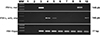
ITS1 sequences were used as targets for designation of the primers and probes to identify L. martiniquensis and L. orientalis/L. chancei and detect trypanosomatids (Table 1, Fig. 1). In the cPCR, the primers, ITS1-L. mar and ITS1-L. ori/cha successfully amplified only the gDNA of L. martiniquensis and L. orientalis/L. chancei, respectively. The ITS1-Tryps primers amplified the gDNA of trypanosomatids (Leishmania spp., T. brucei, and Crithidia sp.) without primer dimerization (File S3). No PCR amplicons of P. vivax, P. falciparum, or M. tuberculosis were observed.
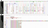 |
Figure 1 Alignment of the conserved regions using Crustal analysis of (A) ITS1 target for Leishmania martiniquensis (green boxes) and ITS1 target for L. orientalis/L. chancei (blue boxes) and (B) ITS1 target for trypanosomatids (red boxes) used to design primers (F, R) and probes (Prob). |
Standard curve of the singleplex and multiplex qPCR assays
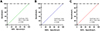
To investigate the detection range of the assays, the DNA standards of L. martiniquensis, L. orientalis, and Crithidia sp. (CLA-KP1) were serially diluted 10-fold and used as templates in the singleplex and multiplex qPCR. The Cq values were plotted against log10 DNA quantity, and standard curves of each target were obtained. No significant differences in sensitivity and specificity in amplifying expected products were observed between the singleplex and multiplex qPCR standard curves. The R2, E, and slope were determined. The R2 of the standard curves was greater than 0.99 and the E values of each target were between 91% and 95%, which were considered acceptable (Fig. 2).
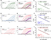 |
Figure 2 Representatives of amplification plots of the singleplex and multiplex qPCR assays and standard curves of the singleplex and multiplex qPCR of L. martiniquensis (A, B, C), L. orientalis (D, E, F, or Crithidia sp. (CLA-KP1) (G, H, I). |
Analytical sensitivity and specificity of the multiplex qPCR assay
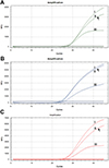
Based on the ROC curve analysis, the cut-off points for L. martiniquensis, L. orientalis, and Crithidia sp. (CLA-KP1) DNA detection were Cq of 38.04, 38.92, and 38.11, respectively (File S4). The LOD was determined through the serial dilutions of known concentrations of L. martiniquensis, L. orientalis, and Crithidia sp. (CLA-KP1) DNA, and the positive detection rates of each dilution were determined. The LOD95 was calculated to be 1.699 fg/reaction (0.0255 parasite equivalents/reaction), 1.717 fg/reaction (0.0292 parasite equivalents/reaction), and 1.763 fg/reaction (0.0882 parasite equivalents/reaction) for L. martiniquensis, L. orientalis, and Crithidia sp. (CLA-KP1), respectively (File S5).
The analytical specificity was assessed by the interference of P. vivax, P. falciparum, and M. tuberculosis on the detection results of the assay. No amplification was found for the DNA of these samples, except for the human DNA in the P. vivax and P. falciparum DNA samples as they were extracted from clinical samples. This result indicates the high analytical specificity of the multiplex qPCR assay (Table 2).
Analytical specificity of the multiplex qPCR assay.
Repeatability and reproducibility of the multiplex qPCR assay
A total of 12 tests of each parasite species were conducted and the mean Cq and %CV values are shown in Table 3. The %CV values of intra- and inter- assays were in the range of 0.23–1.34% and 0.19–2.50%, respectively. The difference between mean Cq values of the intra- and inter-assays was less than 1, suggesting that the multiplex qPCR assay is reliable.
Repeatability and reproducibility analysis of the multiplex RT-qPCR assay.
Evaluation of the multiplex qPCR assay in residual DNA samples
The diagnostic sensitivity and specificity of the multiplex qPCR assay were evaluated using 69 residual DNA samples: 44 positive samples (42 L. martiniquensis and 2 L. orientalis) and 25 negative samples (Table S2). The 42 L. martiniquensis samples and 2 L. orientalis samples were identified by both the multiplex qPCR and the HSP70-I-3′-UTR PCR methods. No false negatives were found, indicating that the multiplex qPCR has a diagnostic sensitivity of 100% for these two species.
For diagnostic specificity, both multiplex qPCR and HSP70-I-3′-UTR PCR methods gave negative results with all 25 negative samples, indicating that this assay has a diagnostic specificity of 100% for L. martiniquensis and L. orientalis. As L. martiniquensis and L. orientalis are trypanosomatids, all 44 positive samples were also true positive for trypanosomatids. The diagnostic sensitivity and specificity of the qPCR assay for detection of trypanosomatids were both 100%. Also, the multiplex qPCR assay demonstrated PPV and NPV of 100% and a perfect agreement (kappa = 1.0) with the reference assay for identification of both Leishmania species and detection of trypanosomatids (Table 4). The multiplex qPCR assay detected parasites ranging from 0.20–1822.01 and 23.33–50.84 parasite equivalents/reaction in the clinical samples of L. martiniquensis and L. orientalis, respectively (Table S2).
Evaluation results of the multiplex qPCR assay compared to the HSP70-I-3′-UTR PCR assay for identification of L. martiniquensis and L. orientalis and detection of trypanosomatid.
Discussion
Most previous studies have focused on developing singleplex qPCR assays for pan-genus Leishmania detection using highly sensitive targets, such as mkDNA or kDNA, spliced-leader (SL) RNA [12, 49], the arginine permease gene (AAP3) [68], HSP70 gene, and 18S rDNA, which characterize the Leishmania genus [12, 55, 71, 73]. In recent years, several qPCR assays have been developed to detect Leishmania spp. DNA in clinical samples. For instance, Eberhardt et al. [9] developed an SL-RNA qPCR assay that demonstrated exceptional analytical sensitivity, detecting 0.005 and 0.002 parasites per mg of liver and spleen tissue, respectively. This SL-RNA qPCR assay is equally effective in detecting L. infantum, L. donovani, L. tropica, L. major, L. mexicana, L. panamensis, L. guyanensis, and L. braziliensis. In another study, qPCR assays targeting the HSP70 and 18S rDNA genes of Leishmania spp. in multiplex with the human RNAse P gene have been developed and validated. The assays can detect up to 0.01 parasite equivalents/reaction and up to 0.1 parasite equivalents/reaction for the HSP70 target. The assays could detect DNA from L. amazonensis, L. guyanensis, L. panamensis, and L. braziliensis [12]. Although these assays have shown excellent sensitivity and specificity for detecting most studied Leishmania spp., no species discrimination is possible.
Here, we successfully developed a novel one-step multiplex qPCR assay that simultaneously identified and quantified L. martiniquensis and L. orientalis/L. chancei parasites and detected and quantified other trypanosomatids in clinical samples, using ITS1 as the molecular target and human RNase P as the internal control gene. The developed assay provided high diagnostic values of 100% sensitivity and high analytical specificity, at a concentration of approximately 1.7 (0.03) fg/reaction (parasite equivalents/reaction) for the LOD95. In addition, the repeatability and reproducibility analysis of the multiplex qPCR assay revealed that the test exhibited good reproducibility across different testing days, with no inconclusive results or statistical differences between replicates. These findings indicate that the assay is reliable for clinical diagnosis and appropriate for clinical application in detection and identification of the new species without the need for parasite isolation and cultivation. In the case of weak positives or near-threshold results, sequencing of the amplified DNA is required to confirm the presence of the target sequence (https://www.epa.gov/sites/default/files/2015-07/documents/epa-qaqc-pcr.pdf).
Recently, the novel duplex TaqMan-based qPCR for the diagnosis of L. martiniquensis and L. orientalis using the ITS1 and the heat shock protein 70 (type I) intergenic region (HSP70-I IR) as targets has demonstrated that the LOD of L. martiniquensis and L. orientalis is approximately 1 copy per reaction [55]. However, this could not be compared to our study, as their study used a standard plasmid as a template for assay analysis. In our study, human DNA was used to dilute the parasite DNA to confirm that no background problem was caused by human DNA in clinical specimens, thereby indicating true sensitivity of our multiplex qPCR assay for diagnosis.
The inability of the developed multiplexed qPCR assay to distinguish between L. orientalis and L. chancei should not limit its clinical utility due to the geographical separation of these species, as L. orientalis is endemic only in Thailand and L. chancei is endemic only in West Africa [20, 25, 26, 33, 60]. However, only two L. orientalis and one L. chancei true-positive samples were available in this study. Therefore, a larger and geographically diverse cohort, including L. orientalis and L. chancei samples from different endemic regions, would be necessary for robust validation in the future.
Like pan-genus Leishmania detection qPCR assays [9, 49], our developed multiplex qPCR assay has a limitation, i.e., that mixed infection of other Leishmania spp. or trypanosomatids cannot be excluded. Since the ITS1-Tryps-Texas Red primer-probe set was designed to detect all trypanosomatids, it could also amplify the DNA of L. martiniquensis, L. orientalis, and L. chancei. Thus, physicians in the synanthropic area where multiple species are endemic should be aware of this limitation. However, the advantage of this multiplex qPCR assay is its ability to screen for other Leishmania spp. and trypanosomatid infections in the same run. Overall, the developed multiplex qPCR assay serves its intended purpose, i.e., identification and quantification of emerging leishmaniasis by L. martiniquensis and L. orientalis/L. chancei parasites and detection and quantification of trypanosomatid infection in humans in a single reaction.
While L. orientalis has only been reported in Thailand [3], the epidemiology of L. martiniquensis reveals a global distribution, with human cases reported in Martinique [8], Thailand (reviewed by [27]), and Myanmar [46], as well as cases in horses in the United States [38, 57], Germany [44], Switzerland [44], and Brazil [37], and in cows in Switzerland [29]. In addition, asymptomatic leishmaniasis cases caused by L. orientalis and L. martiniquensis have been detected in both the northern and southern regions of Thailand [33, 67]. In a southern province in Thailand, an asymptomatic Leishmania infection has been detected among blood donors with a prevalence of 19%, with L. martiniquensis being the predominant species [50]. Investigations regarding whether the asymptomatic individuals could potentially harbor and transmit the parasite through blood products should be conducted, using highly sensitive and reliable methods such as qPCR due to very low blood parasitemia. Furthermore, inspection of the Leishmania infection results, from blood donors, should be performed attentively as gold-standard methods, i.e., Giemsa staining and cultivation cannot be used to identify parasites in asymptomatic carriers [40]. Given the concern over Leishmania transmission via blood transfusion, our developed multiplex qPCR assay provides an additional promising tool for screening blood products for Leishmania DNA. Furthermore, the multiplex qPCR assay would be useful in public health surveillance for the detection of asymptomatic carriers.
Besides the Leishmania parasites, the multiplex qPCR assay can detect trypanosomatids that infect humans, such as T. brucei, facilitating surveillance, monitoring, and management of the diseases. This assay would significantly impact disease management by providing a rapid, accurate, and species-specific diagnosis, thereby enabling prompt treatment and appropriate therapy. The potential spread of Leishmania parasites to non-endemic regions could be due to increased population migration, international travel activities of humans and animal hosts, growth and spread of vector populations, and increased asymptomatic cases [58]. Thus, our developed multiplex qPCR assay could be applied to all endemic areas with leishmaniasis and other trypanosomatid infections worldwide, not limited to Thailand. However, in the future, additional field validation should be performed, exploring the utility of the assay with various clinical samples in different endemic settings, to confirm the actual value of this tool.
Conclusion
The ITS1/human RNase P multiplex qPCR assay for the simultaneous identification and quantification of L. martiniquensis, L. orientalis/L. chancei parasites, and detection and quantification of other trypanosomatids in clinical samples was developed, with high diagnostic values of 100% sensitivity and high specificity. The assay detected a minimum of 0.0255 parasite equivalents/reaction for L. martiniquensis, 0.0292 parasite equivalents/reaction for L. orientalis, and 0.0882 parasite equivalents/reaction for Crithidia sp. (CLA-KP1). This newly developed multiplex qPCR assay offers a rapid, precise, and reliable diagnostic tool for future applications in diagnosing the three new Leishmania species and detecting and quantifying other trypanosomatid parasites. Rapid and effective diagnosis of leishmaniasis would benefit patients to receive appropriate therapy and prompt treatment to reduce possible complications. The developed multiplex qPCR assay would also benefit large-scale screening and surveillance programs in detecting L. martiniquensis, L. orientalis, and trypanosomatid parasites in asymptomatic individuals, especially people living with HIV and blood donors in Thailand and worldwide.
Acknowledgments
We thank Dr. Supaporn Wacharapluesadee, Thai Red Cross Emerging Infectious Diseases Clinical Center, King Chulalongkorn Memorial Hospital, Bangkok, Thailand, for valuable suggestions, Prof. Dr. Somchai Jongwutiwes and Prof. Dr. Chaturong Putaporntip, Department of Parasitology, Faculty of Medicine, Chulalongkorn University, Bangkok, Thailand, for gDNA of P. vivax and P. falciparum and Dr. Suwatchareeporn Rotcheewaphan, Department of Microbiology, Faculty of Medicine, Chulalongkorn University, Bangkok, Thailand, for gDNA of M. tuberculosis.
Funding
This study was funded by the Thailand Science, Research and Innovation Fund Chulalongkorn University (HEA663000030) and the Second Century Fund (C2F), Chulalongkorn University.
Conflicts of interest
The authors declare that they have no competing interests.
Author contribution statement
N.J.: conceptualization; N.J., C.M., P.H., P.P. and S.K.: study design & methodology, investigation & validation; N.J., C.M., P.L., P.S. and A.T.: data analysis; N.J., C.M. and P.L.: writing original draft; N.J., D.G., M.D.U. and P.A.B.: writing – review & editing. All authors read and approved the final version of the manuscript.
Supplementary material
 |
File S1. A. The original version of the primers-probe set of Homo sapiens ribonuclease P (RP forward: AGATTTGGACCTGCGAGCG; RP probe: TTCTGACCTGAAGGCTCTGCGCG; RP reverse: GAGCGGCTGTCTCCACAAGT) designed by Fan et al. [11] is shown in red letters and the modified version (RP-human forward: TCAGCATGGCGGTGTTT; RP-human probe: TTCTGACCTGAAGGCTCTGCGC; RP-human reverse: CGGCTGTCTCCACAAGTC) is in yellow highlight. Both sets were designed from the reference sequence (GenBank accession number): NM_006413.4. B. Results of Blastn of the fragment of 65 bp of the original primers-probe set. C. Results of Blastn of the fragment of 81 bp of the modified version. |
 |
File S2. Representative of optimization for the multiplex qPCR assay using primers-probe concentration for each target at (i) 250 nM primers/100 nM probe, (ii) 125 nM primers/50 nM probe, and (iii) 62.5 nM primers/25 nM probe and 102 fg/reaction for (A) L. martiniquensis, (B) L. orientalis, and (C) Crithidia sp. (CLA-KP1) as templates. The qPCR reactions were conducted according to the recommended thermal cycling protocol: 1 cycle of polymerase activation at 95 °C for 5 min, followed by 45 cycles of PCR at 95 °C for 15 s, and 60 °C for 1 min. |
 |
File S3. PCR products of the ITS1 fragment of 149 bp using ITS1-L. mar primers, the ITS1 fragment of 146 bp using ITS1-L. ori/cha primers, and the ITS1 conserved fragment of 77 bp using ITS1-Tryps primers. Lanes: MW = Molecular weight markers; 1 = Negative control; 2 = human DNA; 3 = L. martiniquensis; 4 = L. orientalis; 5 = L. chancei; 6 = L. infantum; 7 = L. braziliensis; 8 = Crithidia sp. (CLA-KP1); 9 = L. martiniquensis + human DNA; 10 = L. orientalis + human DNA. |
 |
File S4. Receiver operating characteristic (ROC) analysis for cut-off estimation for (A) L. martiniquensis, (B) L. orientalis, and (C) Crithidia sp. (CLA-KP1). |
 |
File S5. Limit of detection (LOD95) of the multiplex qPCR for (A) L. martiniquensis, (B) L. orientalis, and (C) Crithidia sp. (CLA-KP1) was estimated by fitting a logistic regression model with detection results. Each dot indicates an actual detection rate at each dilution. The dashed line in the figure indicates the dilution ratio corresponding to a 95% probability. |
Table S1. Selected sequences of ITS1 targets of trypanosomatids that were used to design the primers and probes in this study. Access Supplementary Material
Table S2. Clinical diagnosis in the 69 residual DNA samples: 44 positive (42 for L. martiniquensis and 2 for L. orientalis) samples extracted from clinical specimens of leishmaniasis patients and 25 negative samples using the multiplex qPCR assay. Access Supplementary Material
References
- Akhoundi M, Downing T, Votýpka J, Kuhls K, Lukeš J, Cannet A, Ravel C, Marty P, Delaunay P, Kasbari M, Granouillac B, Gradoni L, Sereno D. 2017. Leishmania infections: Molecular targets and diagnosis. Molecular Aspects of Medicine, 57, 1–29. [CrossRef] [PubMed] [Google Scholar]
- Alvar J, Aparicio P, Aseffa A, Den Boer M, Cañavate C, Dedet JP, Gradoni L, Ter Horst R, López-Vélez R, Moreno J. 2008. The relationship between leishmaniasis and AIDS: the second 10 years. Clinical Microbiology Reviews, 21, 334–359. [CrossRef] [PubMed] [Google Scholar]
- Anugulruengkitt S, Songtaweesin WN, Thepnarong N, Tangthanapalakul A, Sitthisan M, Chatproedprai S, Wititsuwannakul J, Likitnukul S, Jariyapan N, Weedall GD, Siriyasatien P, Preativatanyou K. 2022. Case report: Simple nodular cutaneous leishmaniasis caused by autochthonous Leishmania (Mundinia) orientalis in an 18-month-old girl: the first pediatric case in Thailand and literature review. American Journal of Tropical Medicine and Hygiene, 108, 44–50. [Google Scholar]
- Asfaram S, Fakhar M, Mohebali M, Ziaei Hezarjaribi H, Mardani A, Ghezelbash B, Akhoundi B, Zarei Z, Moazeni M. 2022. A convenient and sensitive kDNA-PCR for screening of Leishmania infantum latent infection among blood donors in a highly endemic focus, Northwestern Iran. Acta Parasitologica, 67, 842–850. [CrossRef] [PubMed] [Google Scholar]
- Bualert L, Ruang-Areerate T, Mungthin M, Leelayoova S, Siripattanapipong S, Naaglor T, Hongsimakul N, Sroythong S, Rattanalertpaiboon P, Tulpeng P, Piyaraj P. 2024. Incidence and persistence of asymptomatic Leishmania infection among HIV-infected patients in Trang province, Southern Thailand: a cohort study. PLoS Neglected Tropical Diseases, 18, e0012581. [CrossRef] [PubMed] [Google Scholar]
- Carbonara M, Mendoza-Roldan JA, Bezerra-Santos MA, de Abreu Teles PP, Lia RP, Locantore F, Iatta R, Volf P, Otranto D. 2024. Leishmania spp. in equids and their potential vectors in endemic areas of canine leishmaniasis. PLoS Neglected Tropical Diseases, 18, e0012290. [CrossRef] [PubMed] [Google Scholar]
- Çorbacıoğlu ŞK, Aksel G. 2023. Receiver operating characteristic curve analysis in diagnostic accuracy studies: a guide to interpreting the area under the curve value. Turkish Journal of Emergency Medicine, 23, 195–198. [CrossRef] [PubMed] [Google Scholar]
- Desbois N, Pratlong F, Quist D, Dedet JP. 2014. Leishmania (Leishmania) martiniquensis n. sp. (Kinetoplastida: Trypanosomatidae), description of the parasite responsible for cutaneous leishmaniasis in Martinique Island (French West Indies). Parasite, 21, 12. [CrossRef] [EDP Sciences] [PubMed] [Google Scholar]
- Eberhardt E, Van den Kerkhof M, Bulté D, Mabille D, Van Bockstal L, Monnerat S, Alves F, Mbui J, Delputte P, Cos P, Hendrickx S, Maes L, Caljon G. 2018. Evaluation of a pan-Leishmania spliced-leader RNA detection method in human blood and experimentally infected syrian golden hamsters. Journal of Molecular Diagnostics, 20, 253–263. [CrossRef] [Google Scholar]
- Escobar TA, Dowich G, Dos Santos TP, Zuravski L, Duarte CA, Lübeck I, Manfredini V. 2019. Assessment of Leishmania infantum infection in equine populations in a canine visceral leishmaniosis transmission area. BMC Veterinary Research, 15, 381. [CrossRef] [PubMed] [Google Scholar]
- Fan J, Cui D, Lau S, Xie G, Guo X, Zheng S, Huang X, Yang S, Yang X, Huo Z, Yu F, Lou J, Tian L, Li X, Dong Y, Zhu Q, Chen Y. 2014. Detection of a novel avian influenza A (H7N9) virus in humans by multiplex one-step real-time RT-PCR assay. BMC Infectious Diseases, 14, 541. [CrossRef] [PubMed] [Google Scholar]
- Filgueira CPB, Moreira OC, Cantanhêde LM, de Farias HMT, Porrozzi R, Britto C, Boité MC, Cupolillo E. 2020. Comparison and clinical validation of qPCR assays targeting Leishmania 18S rDNA and HSP70 genes in patients with American Tegumentary Leishmaniasis. PLoS Neglected Tropical Diseases, 14, e0008750. [CrossRef] [PubMed] [Google Scholar]
- Galluzzi L, Ceccarelli M, Diotallevi A, Menotta M, Magnani M. 2018. Real-time PCR applications for diagnosis of leishmaniasis. Parasites & Vectors, 11, 273. [CrossRef] [PubMed] [Google Scholar]
- Ghobakhloo N, Motazedian MH, Naderi S, Ebrahimi S. 2019. Isolation of Crithidia spp. from lesions of immunocompetent patients with suspected cutaneous leishmaniasis in Iran. Tropical Medicine and International Health, 24, 116–126. [CrossRef] [PubMed] [Google Scholar]
- Ghosh S, Banerjee P, Sarkar A, Datta S, Chatterjee M. 2012. Coinfection of Leptomonas seymouri and Leishmania donovani in Indian leishmaniasis. Journal of Clinical Microbiology, 50, 2774–2778. [CrossRef] [PubMed] [Google Scholar]
- Gow I, Smith NC, Stark D, Ellis J. 2022. Laboratory diagnostics for human Leishmania infections: a polymerase chain reaction-focussed review of detection and identification methods. Parasites & Vectors, 15, 412. [CrossRef] [PubMed] [Google Scholar]
- Ibarra-Meneses AV, Corbeil A, Wagner V, Onwuchekwa C, Fernandez-Prada C. 2022. Identification of asymptomatic Leishmania infections: a scoping review. Parasites & Vectors, 15, 5. [CrossRef] [PubMed] [Google Scholar]
- Inga R, De Doncker S, Gomez J, Lopez M, Garcia R, Le Ray D, Arevalo J, Dujardin JC. 1998. Relation between variation in copy number of ribosomal RNA encoding genes and size of harbouring chromosomes in Leishmania of subgenus Viannia. Molecular and Biochemical Parasitology, 92, 219–228. [CrossRef] [PubMed] [Google Scholar]
- Jariyapan N, Bates MD, Bates PA. 2021. Molecular identification of two newly identified human pathogens causing leishmaniasis using PCR-based methods on the 3’ untranslated region of the heat shock protein 70 (type I) gene. PLoS Neglected Tropical Diseases, 15, e0009982. [CrossRef] [PubMed] [Google Scholar]
- Jariyapan N, Daroontum T, Jaiwong K, Chanmol W, Intakhan N, Sor-Suwan S, Siriyasatien P, Somboon P, Bates MD, Bates PA. 2018. Leishmania (Mundinia) orientalis n. sp. (Trypanosomatidae), a parasite from Thailand responsible for localised cutaneous leishmaniasis. Parasites & Vectors, 11, 351. [CrossRef] [PubMed] [Google Scholar]
- Jariyapan N, Dissook S, Noisagul P, Thongkumkoon P, Mano C, Kittichaiworakul R, Junkum A, Tantiworawit A, Pescher P, Späth GF, Almutairi H, Siriyasatien P. 2025. Genome analyses of amphotericin B-susceptible and -resistant strains of Leishmania (Mundinia) martiniquensis reveal variations potentially related to amphotericin B resistance. Current Research in Parasitology & Vector-Borne Diseases, 7, 100255. [CrossRef] [PubMed] [Google Scholar]
- Kaewmee S, Mano C, Phanitchakun T, Ampol R, Yasanga T, Pattanawong U, Junkum A, Siriyasatien P, Bates PA, Jariyapan N. 2023. Natural infection with Leishmania (Mundinia) martiniquensis supports Culicoides peregrinus (Diptera: Ceratopogonidae) as a potential vector of leishmaniasis and characterization of a Crithidia sp. isolated from the midges. Frontiers in Microbiology, 14, 1235254. [CrossRef] [PubMed] [Google Scholar]
- Klymus KE, Merkes CM, Allison MJ, Goldberg CS, Helbing CC, Hunter ME, Jackson CA, Lance RF, Mangan AM, Monroe EM, Piaggio AJ, Stokdyk JP, Wilson CC, Richter CA. 2020. Reporting the limits of detection and quantification for environmental DNA assays. Environmental DNA, 2, 271–282. [CrossRef] [Google Scholar]
- Kostygov AY, Butenko A, Yurchenko V. 2019. On monoxenous trypanosomatids from lesions of immunocompetent patients with suspected cutaneous leishmaniasis in Iran. Tropical Medicine and International Health, 24, 127–128. [CrossRef] [PubMed] [Google Scholar]
- Kwakye-Nuako G, Mosore MT, Boakye D, Bates PA. 2023. Description, biology, and medical significance of Leishmania (Mundinia) chancei n. sp. (Kinetoplastea: Trypanosomatidae) from Ghana and Leishmania (Mundinia) procaviensis n. sp. (Kinetoplastea: Trypanosomatidae) from Namibia. Journal of Parasitology, 109, 43–50. [CrossRef] [PubMed] [Google Scholar]
- Kwakye-Nuako G, Mosore MT, Duplessis C, Bates MD, Puplampu N, Mensah-Attipoe I, Desewu K, Afegbe G, Asmah RH, Jamjoom MB, Ayeh-Kumi PF, Boakye DA, Bates PA. 2015. First isolation of a new species of Leishmania responsible for human cutaneous leishmaniasis in Ghana and classification in the Leishmania enriettii complex. International Journal for Parasitology, 45, 679–684. [CrossRef] [PubMed] [Google Scholar]
- Leelayoova S, Siripattanapipong S, Manomat J, Piyaraj P, Tan-Ariya P, Bualert L, Mungthin M. 2017. Leishmaniasis in Thailand: A review of causative agents and situations. American Journal of Tropical Medicine and Hygiene, 96, 534–542. [Google Scholar]
- Leon W, Fouts DL, Manning J. 1978. Sequence arrangement of the 16S and 26S rRNA genes in the pathogenic haemoflagellate Leishmania donovani. Nucleic Acids Research, 5, 491–504. [CrossRef] [PubMed] [Google Scholar]
- Lobsiger L, Müller N, Schweizer T, Frey CF, Wiederkehr D, Zumkehr B, Gottstein B. 2011. An autochthonous case of cutaneous bovine leishmaniasis in Switzerland. Veterinary Parasitology, 169, 408–414. [Google Scholar]
- Maharom P, Siripattanapipong S, Mungthin M, Naaglor T, Sukkawee R, Pudkorn R, Wattana W, Wanachiwanawin D, Areechokchai D, Leelayoova S. 2008. Visceral leishmaniasis caused by Leishmania infantum in Thailand. Southeast Asian Journal of Tropical Medicine and Public Health, 39, 988–990. [Google Scholar]
- Mano C, Kongkaew A, Tippawangkosol P, Junkum A, Siriyasatien P, Jariyapan N. 2023. In vitro susceptibility to miltefosine of amphotericin B-resistant Leishmania (Mundinia) martiniquensis. Parasitology Research, 122, 3027–3035. [CrossRef] [PubMed] [Google Scholar]
- Mano C, Kongkaew A, Tippawangkosol P, Somboon P, Roytrakul S, Pescher P, Späth GF, Uthaipibull C, Tantiworawit A, Siriyasatien P, Jariyapan N. 2023. Amphotericin B resistance correlates with increased fitness in vitro and in vivo in Leishmania (Mundinia) martiniquensis. Frontiers in Microbiology, 14, 1156061. [CrossRef] [PubMed] [Google Scholar]
- Manomat J, Leelayoova S, Bualert L, Tan-Ariya P, Siripattanapipong S, Mungthin M, Naaglor T, Piyaraj P. 2017. Prevalence and risk factors associated with Leishmania infection in Trang Province, southern Thailand. PLoS Neglected Tropical Diseases, 11, e0006095. [CrossRef] [PubMed] [Google Scholar]
- Martín-Escolano J, Marín C, Rosales MJ, Tsaousis AD, Medina-Carmona E, Martín-Escolano R. 2022. An updated view of the Trypanosoma cruzi life cycle: intervention points for an effective treatment. ACS Infectious Diseases, 8, 1107–1115. [CrossRef] [PubMed] [Google Scholar]
- Maruyama SR, de Santana AKM, Takamiya NT, Takahashi TY, Rogerio LA, Oliveira CAB, Milanezi CM, Trombela VA, Cruz AK, Jesus AR, Barreto AS, da Silva AM, Almeida RP, Ribeiro JM, Silva JS. 2019. Non-Leishmania parasite in fatal visceral leishmaniasis-like disease, Brazil. Emerging Infectious Diseases, 25, 2088–2092. [CrossRef] [PubMed] [Google Scholar]
- McHugh ML. 2012. Interrater reliability: the kappa statistic. Biochemia Medica, 22, 276–282. [CrossRef] [PubMed] [Google Scholar]
- Mendes Junior AAV, Filgueira CPB, Miranda LFC, de Almeida AB, Cantanhêde LM, Fagundes A, Pereira SA, Menezes RC, Cupolillo E. 2023. First report of Leishmania (Mundinia) martiniquensis in South American territory and confirmation of Leishbunyavirus infecting this parasite in a mare. Memórias do Instituto Oswaldo Cruz, 118, e220220. [CrossRef] [PubMed] [Google Scholar]
- Menezes RC, Campos MP, Popielarczyk M, Kiupel M. 2019. Cutaneous Leishmaniosis caused by Leishmania martiniquensis in a horse in Florida. Journal of Comparative Pathology, 173, 13–18. [CrossRef] [PubMed] [Google Scholar]
- Mock DJ, Hollenbaugh JA, Daddacha W, Overstreet MG, Lazarski CA, Fowell DJ, Kim B. 2012. Leishmania induces survival, proliferation and elevated cellular dNTP levels in human monocytes promoting acceleration of HIV co-infection. PLoS Pathogens, 8, e1002635. [CrossRef] [PubMed] [Google Scholar]
- Mohammed R, Melkamu R, Pareyn M, Abdellati S, Bogale T, Engidaw A, Kinfu A, Girma T, van Griensven J. 2023. Detection of asymptomatic Leishmania infection in blood donors at two blood banks in Ethiopia. PLoS Neglected Tropical Diseases, 17, e0011142. [CrossRef] [PubMed] [Google Scholar]
- Monaghan TF, Rahman SN, Agudelo CW, Wein AJ, Lazar JM, Everaert K, Dmochowski RR. 2021. Foundational statistical principles in medical research: sensitivity, specificity, positive predictive value, and negative predictive value. Medicina (Kaunas, Lithuania), 57, 503. [CrossRef] [PubMed] [Google Scholar]
- Morales-Yuste M, Martín-Sánchez J, Corpas-Lopez V. 2022. Canine leishmaniasis: update on epidemiology, diagnosis, treatment, and prevention. Veterinary Sciences, 9, 387. [CrossRef] [PubMed] [Google Scholar]
- Mota TF, Brodskyn CI, Morello LG, Marchini FK, Krieger MA, de Cássia Pontello Rampazzo R, Fraga DBM. 2022. Multiplex qPCR assay to determine Leishmania infantum load in Lutzomyia longipalpis sandfly samples. Medical and Veterinary Entomology, 36, 176–184. [CrossRef] [PubMed] [Google Scholar]
- Müller N, Welle M, Lobsiger L, Stoffel MH, Boghenbor KK, Hilbe M, Gottstein B, Frey CF, Geyer C, von Bomhard W. 2009. Occurrence of Leishmania sp. in cutaneous lesions of horses in Central Europe. Veterinary Parasitology, 166, 346–351. [CrossRef] [PubMed] [Google Scholar]
- Ni M, Xu H, Luo J, Liu W, Zhou D. 2021. Simultaneous detection and differentiation of SARS-CoV-2, influenza A virus and influenza B virus by one-step quadruplex real-time RT-PCR in patients with clinical manifestations. International Journal of Infectious Diseases, 103, 517–524. [CrossRef] [Google Scholar]
- Noppakun N, Kraivichian K, Siriyasatien P. 2014. Disseminated dermal leishmaniasis caused by Leishmania siamensis in a systemic steroid therapy patient. American Journal of Tropical Medicine and Hygiene, 91, 869–870. [CrossRef] [PubMed] [Google Scholar]
- Noyes HA, Stevens JR, Teixeira M, Phelan J, Holz P. 1999. A nested PCR for the ssrRNA gene detects Trypanosoma binneyi in the platypus and Trypanosoma sp. in wombats and kangaroos in Australia. International Journal for Parasitology, 29, 331–339. [CrossRef] [PubMed] [Google Scholar]
- Panahi E, Stanisic DI, Skinner EB, Faddy HM, Young MK, Herrero LJ. 2023. Detection of Leishmania (Mundinia) macropodum (Kinetoplastida: Trypanosomatidae) and heterologous Leishmania species antibodies among blood donors in a region of Australia with marsupial Leishmania endemicity. International Journal of Infectious Diseases, 130, 42–47. [CrossRef] [Google Scholar]
- Pareyn M, Hendrickx R, Girma N, Hendrickx S, Van Bockstal L, Van Houtte N, Shibru S, Maes L, Leirs H, Caljon G. 2020. Evaluation of a pan-Leishmania SL RNA qPCR assay for parasite detection in laboratory-reared and field-collected sand flies and reservoir hosts. Parasites & Vectors, 13, 276. [CrossRef] [PubMed] [Google Scholar]
- Piyaraj P, Bualert L, Kalrat A, Leelayoova S, Ruang-Areerate T, Theprin N, Naaglor T, Mungthin M. 2024. Asymptomatic Leishmania infection among blood donors in a southern province of Thailand. American Journal of Tropical Medicine and Hygiene, 111, 804–813. [Google Scholar]
- Ponte-Sucre A. 2016. An overview of Trypanosoma brucei infections: An intense host-parasite interaction. Frontiers in Microbiology, 7, 2126. [CrossRef] [PubMed] [Google Scholar]
- Pothirat T, Tantiworawit A, Chaiwarith R, Jariyapan N, Wannasan A, Siriyasatien P, Supparatpinyo K, Bates MD, Kwakye-Nuako G, Bates PA. 2014. First isolation of Leishmania from Northern Thailand: case report, identification as Leishmania martiniquensis and phylogenetic position within the Leishmania enriettii complex. PLoS Neglected Tropical Diseases, 8, e3339. [CrossRef] [PubMed] [Google Scholar]
- Preativatanyou K, Chinwirunsirisup K, Phumee A, Khositharattanakool P, Sunantaraporn S, Depaquit J, Siriyasatien P. 2023. Species diversity of phlebotomine sand flies and sympatric occurrence of Leishmania (Mundinia) martiniquensis, Leishmania (Leishmania) donovani complex, and Trypanosoma spp. in the visceral leishmaniasis focus of southern Thailand. Acta Tropica, 244, 106949. [CrossRef] [PubMed] [Google Scholar]
- Preativatanyou K, Songumpai N, Khositharattanakool P, Ampol R, Promrangsee C, Sricharoensuk C, Phadungsaksawasdi K, Pataradool T, Becvar T, Vojtkova B, Volf P, Siriyasatien P. 2024. Novel duplex TaqMan-based quantitative PCR for rapid and accurate diagnosis of Leishmania (Mundinia) martiniquensis and Leishmania (Mundinia) orientalis, responsible for autochthonous leishmaniasis in Thailand. Current Research in Parasitology and Vector-Borne Diseases, 6, 100217. [CrossRef] [Google Scholar]
- Ramírez JD, Cao L, Castillo-Castañeda AC, Patino LH, Ayala MS, Cordon-Cardo C, Sordillo EM, Paniz-Mondolfi A. 2023. Clinical performance of a quantitative pan-genus Leishmania real-time PCR assay for diagnosis of cutaneous and visceral leishmaniasis. Practical Laboratory Medicine, 37, e00341. [CrossRef] [PubMed] [Google Scholar]
- Requena JM, Chicharro C, García L, Parrado R, Puerta CJ, Cañavate C. 2012. Sequence analysis of the 3’-untranslated region of HSP70 (type I) genes in the genus Leishmania: its usefulness as a molecular marker for species identification. Parasites & Vectors, 5, 87. [CrossRef] [PubMed] [Google Scholar]
- Reuss SM, Dunbar MD, Calderwood Mays MB, Owen JL, Mallicote MF, Archer LL, Wellehan JFJr. 2012. Autochthonous Leishmania siamensis in horse, Florida, USA. Emerging Infectious Diseases, 18, 1545–1547. [CrossRef] [PubMed] [Google Scholar]
- Riebenbauer K, Czerny S, Egg M, Urban N, Kinaciyan T, Hampel A, Fidelsberger L, Karlhofer F, Porkert S, Walochnik J, Handisurya A. 2024. The changing epidemiology of human leishmaniasis in the non-endemic country of Austria between 2000 to 2021, including a congenital case. PLoS Neglected Tropical Diseases, 18, e0011875. [CrossRef] [PubMed] [Google Scholar]
- Rogerio LA, Takahashi TY, Cardoso L, Takamiya NT, de Melo EV, de Jesus AR, de Oliveira FA, Forrester S, Jeffares DC, da Silva JS, Ribeiro JM, Almeida RP, Maruyama SR. 2023. Co-infection of Leishmania infantum and a Crithidia-related species in a case of refractory relapsed visceral leishmaniasis with non-ulcerated cutaneous manifestation in Brazil. International Journal of Infectious Diseases, 133, 85–88. [CrossRef] [Google Scholar]
- Ruang-Areerate T, Ruang-Areerate P, Manomat J, Naaglor T, Piyaraj P, Mungthin M, Leelayoova S, Siripattanapipong S. 2023. Genetic variation and geographic distribution of Leishmania orientalis and Leishmania martiniquensis among Leishmania/HIV co-infection in Thailand. Scientific Reports, 13, 23094. [CrossRef] [PubMed] [Google Scholar]
- Sales KGDS, Miranda DEO, Paiva MHS, Figueredo LA, Otranto D, Dantas-Torres F. 2020. Fast multiplex real-time PCR assay for simultaneous detection of dog and human blood and Leishmania parasites in sand flies. Parasites & Vectors, 13, 131. [CrossRef] [PubMed] [Google Scholar]
- Sapp SGH, Low R, Nine G, Nascimento FS, Qvarnstrom Y, Barratt JLN. 2024. Genetic characterization and description of Leishmania (Leishmania) ellisi sp. nov.: a new human-infecting species from the USA. Parasitology Research, 123, 52. [CrossRef] [Google Scholar]
- Sarasombath PT. 2018. Leishmaniasis: An evolving public health concern in Thailand. Siriraj Medical Journal, 70, 363–376. [Google Scholar]
- Silva LP, Montenegro S, Werkauser R, Sales KGDS, Soares FCS, Costa VMA, Bezerra AC, Pinto MBDA, Ferreira SM, Neitzke-Abreu HC, Dantas-Torres F, Lima Junior MSDC. 2020. Asymptomatic Leishmania infection in blood donors from a major blood bank in Northeastern Brazil: a cross-sectional study. Revista do Instituto de Medicina Tropical de São Paulo, 62, e92. [CrossRef] [Google Scholar]
- Singh OP, Hasker E, Sacks D, Boelaert M, Sundar S. 2014. Asymptomatic Leishmania infection: a new challenge for Leishmania control. Clinical Infectious Diseases, 58, 1424–1429. [CrossRef] [PubMed] [Google Scholar]
- Srivarasat S, Brownell N, Siriyasatien P, Noppakun N, Asawanonda P, Rattanakorn K, Preativatanyou K, Kumtornrut C. 2022. Case report: autochthonous disseminated cutaneous, mucocutaneous, and visceral leishmaniasis caused by Leishmania martiniquensis in a patient with HIV/AIDS from northern Thailand and literature review. American Journal of Tropical Medicine and Hygiene, 107, 1196–1202. [CrossRef] [PubMed] [Google Scholar]
- Sriwongpan P, Nedsuwan S, Manomat J, Charoensakulchai S, Lacharojana K, Sankwan J, Kobpungton N, Sriwongpun T, Leelayoova S, Mungthin M, Siripattanapipong S, Ruang-Areerate T, Naaglor T, Eamchotchawalit T, Piyaraj P. 2021. Prevalence and associated risk factors of Leishmania infection among immunocompetent hosts, a community-based study in Chiang Rai, Thailand. PLoS Neglected Tropical Diseases, 15, e0009545. [CrossRef] [PubMed] [Google Scholar]
- Tellevik MG, Muller KE, Løkken KR, Nerland AH. 2014. Detection of a broad range of Leishmania species and determination of parasite load of infected mouse by real-time PCR targeting the arginine permease gene AAP3. Acta Tropica, 137, 99–104. [CrossRef] [PubMed] [Google Scholar]
- Thakur L, Kushwaha HR, Negi A, Jain A, Jain M. 2020. Leptomonas seymouri co-infection in cutaneous leishmaniasis cases caused by Leishmania donovani from Himachal Pradesh, India. Frontiers in Cellular and Infection Microbiology, 10, 345. [CrossRef] [PubMed] [Google Scholar]
- Villalba E, Ramírez JL. 1982. Ribosomal DNA of Leishmania brasiliensis: number of ribosomal copies and gene isolation. Journal of Protozoology, 29, 438–441. [CrossRef] [PubMed] [Google Scholar]
- Weirather JL, Jeronimo SM, Gautam S, Sundar S, Kang M, Kurtz MA, Haque R, Schriefer A, Talhari S, Carvalho EM, Donelson JE, Wilson ME. 2011. Serial quantitative PCR assay for detection, species discrimination, and quantification of Leishmania spp. in human samples. Journal of Clinical Microbiology, 49, 3892–3904. [CrossRef] [PubMed] [Google Scholar]
- World Health Organization. 2023. Fact sheets: Leishmaniasis. Available at https://www.who.int/news-room/fact-sheets/detail/leishmaniasis (accessed November 31, 2024). [Google Scholar]
- Wu Y, Tian X, Song N, Huang M, Wu Z, Li S, Waterfield NR, Zhan B, Wang L, Yang G. 2020. Application of quantitative PCR in the diagnosis and evaluating treatment efficacy of leishmaniasis. Frontiers in Cellular and Infection Microbiology, 10, 581639. [CrossRef] [PubMed] [Google Scholar]
Cite this article as: Mano C, Hirunpatrawong P, Prasertsilp P, Kaewmee S, Limprasert P, Siriyasatien P, Tantiworawit A, Gatherer D, Urbaniak MD, Bates PA & Jariyapan N. 2025. A one-step multiplex qPCR assay for simultaneous identification and quantification of Leishmania martiniquensis and Leishmania orientalis/Leishmania chancei and detection and quantification of trypanosomatids in clinical samples. Parasite 32, 37. https://doi.org/10.1051/parasite/2025030.
All Tables
Primers and probes for the ITS1 and human RNase P targets used in the developed multiplex qPCR assay.
Evaluation results of the multiplex qPCR assay compared to the HSP70-I-3′-UTR PCR assay for identification of L. martiniquensis and L. orientalis and detection of trypanosomatid.
All Figures
|
Figure 1 Alignment of the conserved regions using Crustal analysis of (A) ITS1 target for Leishmania martiniquensis (green boxes) and ITS1 target for L. orientalis/L. chancei (blue boxes) and (B) ITS1 target for trypanosomatids (red boxes) used to design primers (F, R) and probes (Prob). |
| In the text | |
|
Figure 2 Representatives of amplification plots of the singleplex and multiplex qPCR assays and standard curves of the singleplex and multiplex qPCR of L. martiniquensis (A, B, C), L. orientalis (D, E, F, or Crithidia sp. (CLA-KP1) (G, H, I). |
| In the text | |
|
File S1. A. The original version of the primers-probe set of Homo sapiens ribonuclease P (RP forward: AGATTTGGACCTGCGAGCG; RP probe: TTCTGACCTGAAGGCTCTGCGCG; RP reverse: GAGCGGCTGTCTCCACAAGT) designed by Fan et al. [11] is shown in red letters and the modified version (RP-human forward: TCAGCATGGCGGTGTTT; RP-human probe: TTCTGACCTGAAGGCTCTGCGC; RP-human reverse: CGGCTGTCTCCACAAGTC) is in yellow highlight. Both sets were designed from the reference sequence (GenBank accession number): NM_006413.4. B. Results of Blastn of the fragment of 65 bp of the original primers-probe set. C. Results of Blastn of the fragment of 81 bp of the modified version. |
| In the text | |
|
File S2. Representative of optimization for the multiplex qPCR assay using primers-probe concentration for each target at (i) 250 nM primers/100 nM probe, (ii) 125 nM primers/50 nM probe, and (iii) 62.5 nM primers/25 nM probe and 102 fg/reaction for (A) L. martiniquensis, (B) L. orientalis, and (C) Crithidia sp. (CLA-KP1) as templates. The qPCR reactions were conducted according to the recommended thermal cycling protocol: 1 cycle of polymerase activation at 95 °C for 5 min, followed by 45 cycles of PCR at 95 °C for 15 s, and 60 °C for 1 min. |
| In the text | |
|
File S3. PCR products of the ITS1 fragment of 149 bp using ITS1-L. mar primers, the ITS1 fragment of 146 bp using ITS1-L. ori/cha primers, and the ITS1 conserved fragment of 77 bp using ITS1-Tryps primers. Lanes: MW = Molecular weight markers; 1 = Negative control; 2 = human DNA; 3 = L. martiniquensis; 4 = L. orientalis; 5 = L. chancei; 6 = L. infantum; 7 = L. braziliensis; 8 = Crithidia sp. (CLA-KP1); 9 = L. martiniquensis + human DNA; 10 = L. orientalis + human DNA. |
| In the text | |
|
File S4. Receiver operating characteristic (ROC) analysis for cut-off estimation for (A) L. martiniquensis, (B) L. orientalis, and (C) Crithidia sp. (CLA-KP1). |
| In the text | |
|
File S5. Limit of detection (LOD95) of the multiplex qPCR for (A) L. martiniquensis, (B) L. orientalis, and (C) Crithidia sp. (CLA-KP1) was estimated by fitting a logistic regression model with detection results. Each dot indicates an actual detection rate at each dilution. The dashed line in the figure indicates the dilution ratio corresponding to a 95% probability. |
| In the text | |
Current usage metrics show cumulative count of Article Views (full-text article views including HTML views, PDF and ePub downloads, according to the available data) and Abstracts Views on Vision4Press platform.
Data correspond to usage on the plateform after 2015. The current usage metrics is available 48-96 hours after online publication and is updated daily on week days.
Initial download of the metrics may take a while.