| Issue |
Parasite
Volume 32, 2025
|
|
|---|---|---|
| Article Number | 64 | |
| Number of page(s) | 13 | |
| DOI | https://doi.org/10.1051/parasite/2025057 | |
| Published online | 29 September 2025 | |
Research Article
Molecular analysis of DHFR and DHPS gene mutations in Plasmodium cynomolgi from humans and macaques in Southeast Asia
Analyse moléculaire des mutations des gènes DHFR et DHPS chez Plasmodium cynomolgi chez l’homme et le macaque en Asie du Sud-Est
1
Department of Molecular Tropical Medicine and Genetics, Faculty of Tropical Medicine, Mahidol University, Bangkok 10400, Thailand
2
Department of Social and Applied Science, College of Industrial Technology, King Mongkut’s University of Technology North Bangkok, Bangkok 10800, Thailand
3
Department of Biochemistry, Faculty of Science, Kasetsart University, Bangkok 10903, Thailand
4
National Primate Research Center of Thailand, Chulalongkorn University, Saraburi 18110, Thailand
5
Department of Biology, Faculty of Science, Chulalongkorn University, Bangkok 10330, Thailand
6
Mahidol-Oxford Tropical Medicine Research Unit, Faculty of Tropical Medicine, Mahidol University, Bangkok 10400, Thailand
7
Centre for Tropical Medicine and Global Health, Nuffield Department of Medicine, University of Oxford, Old Road Campus, Oxford OX3 7LF, UK
* Corresponding author: This email address is being protected from spambots. You need JavaScript enabled to view it.
; This email address is being protected from spambots. You need JavaScript enabled to view it.
Received:
1
January
2025
Accepted:
6
September
2025
Abstract
Plasmodium cynomolgi is an emerging zoonotic malaria parasite in Southeast Asia, infecting both humans and macaques. In this study, we investigated mutations in the DHFR and DHPS genes of P. cynomolgi from humans and macaques, comparing them to known resistance mutations in P. falciparum and P. vivax. We also examined how these mutations affect antifolate drug binding, which may influence treatment efficacy and resistance. Nine asymptomatic human blood samples from Cambodia and 29 macaque samples from Thailand were analyzed. Human samples included eight P. cynomolgi monoinfections and one mixed infection with P. vivax, while all macaque samples were monoinfections. The PcyDHFR and PcyDHPS genes were amplified, sequenced, and subjected to haplotype analysis. Human samples from Battambang, Cambodia were 100% identical to the P. cynomolgi RO strain, showing no DHFR mutations and one DHPS mutation (V451I). In contrast, macaque samples from Saraburi, Thailand showed PcyDHFR mutations N44T and C49S, and two haplotypes based on I7 variation – haplotype 1 (72.41%) with wild-type I7 and haplotype 2 (27.59%) with the I7 mutation. PcyDHPS mutations were identical across macaque isolates. Protein structures of PcyDHFR and PcyDHPS were modeled using SWISS-MODEL, focusing on the N- and C-terminals. Mutations occurred near catalytic sites but did not significantly affect binding affinity, based on molecular docking with eight antifolate drugs. These findings suggest that current antifolate drugs remain potentially effective against P. cynomolgi, and highlight the importance of monitoring drug resistance in zoonotic malaria.
Résumé
Plasmodium cynomolgi est un parasite zoonotique du paludisme émergent en Asie du Sud-Est, infectant à la fois l’homme et le macaque. Cette étude a examiné les mutations des gènes DHFR et DHPS de P. cynomolgi chez l’homme et le macaque, en les comparant aux mutations de résistance connues chez P. falciparum et P. vivax. Nous avons également examiné l’impact de ces mutations sur la liaison aux antifolates, ce qui pourrait influencer l’efficacité et la résistance au traitement. Neuf échantillons de sang humain asymptomatique provenant du Cambodge et 29 échantillons de sang de macaques provenant de Thaïlande ont été analysés. Les échantillons humains comprenaient huit mono-infections à P. cynomolgi et une infection mixte à P. vivax, tandis que tous les échantillons de macaques étaient des mono-infections. Les gènes PcyDHFR et PcyDHPS ont été amplifiés, séquencés et soumis à une analyse d’haplotype. Les échantillons humains de Battambang, au Cambodge, étaient identiques à 100 % à la souche RO de P. cynomolgi, ne présentant aucune mutation dhfr et une mutation dhps (V451I). En revanche, les échantillons issus de macaques de Saraburi, en Thaïlande, présentaient les mutations N44T et C49S du gène PcyDHFR, ainsi que deux haplotypes basés sur la variation I7 : l’haplotype 1 (72,41 %) avec le gène I7 sauvage et l’haplotype 2 (27,59 %) avec la mutation I7. Les mutations du gène PcyDHPS étaient identiques entre les isolats de macaques. Les structures protéiques de PcyDHFR et de PcyDHPS ont été modélisées à l’aide de SWISS-MODEL, en se concentrant sur les extrémités N et C. Les mutations se sont produites à proximité des sites catalytiques, mais n’ont pas eu d’effet significatif sur l’affinité de liaison, d’après l’amarrage moléculaire avec huit médicaments antifolates. Ces résultats suggèrent que les médicaments antifolates actuels restent potentiellement efficaces contre P. cynomolgi et soulignent l’importance de surveiller la résistance aux médicaments dans le paludisme zoonotique.
Key words: Plasmodium cynomolgi / Pcydhfr / Pcydhps / Simian malaria
Edited by: Jean-Lou Justine.
© R. Sangsri et al., published by EDP Sciences, 2025
 This is an Open Access article distributed under the terms of the Creative Commons Attribution License (https://creativecommons.org/licenses/by/4.0), which permits unrestricted use, distribution, and reproduction in any medium, provided the original work is properly cited.
Introduction
Malaria is a major global health concern, particularly in tropical and subtropical regions, where over 200 million cases are reported annually. While Plasmodium falciparum and Plasmodium vivax are the most well-known species affecting humans, recent attention has turned toward zoonotic malaria, specifically infections caused by P. cynomolgi, a parasite that primarily infects macaques. Evidence has emerged of its ability to infect humans, although infections are often asymptomatic or mild. Nevertheless, it is a potential emerging zoonotic threat, especially in Southeast Asia, where human populations areas frequently overlap with primate habitats [16, 19]. In Cambodia and Thailand, both human and macaque populations carry P. cynomolgi; thus, these regions are key areas for studying the zoonotic transmission dynamics of malaria [37]. Given the increasing proximity between humans and wildlife due to habitat encroachment, closely monitoring these zoonotic pathogens is critical. Furthermore, as drug-resistant strains of malaria parasites become more prevalent, understanding the genetic mutations in P. cynomolgi that contribute to drug resistance is essential for developing future therapeutic strategies [26].
The dihydrofolate reductase (DHFR) and dihydropteroate synthase (DHPS) genes play pivotal roles in the development of antifolate drug resistance in malaria parasites, including P. cynomolgi. The DHFR gene encodes an enzyme that reduces dihydrofolate to tetrahydrofolate, which is essential for DNA synthesis. Mutations in DHFR, such as N51I, C59R, S108N, and I164L, reduce the effectiveness of antifolate drugs such as pyrimethamine by altering the enzyme’s binding site in a manner that enables drug resistance [7]. Similarly, in the DHPS gene (which codes for an enzyme involved in folate biosynthesis), mutations at key positions, such as A437G and K540E, disrupt drug binding and confer resistance to sulfa drugs such as sulfadoxine and sulfamethoxazole [33]. Drug resistance due to these mutations is particularly concerning, because it can lead to treatment failure with sulfadoxine-pyrimethamine (SP) combination therapy, as has been seen in human malaria parasites [26]. Moreover, these mutations in P. cynomolgi raise concerns about zoonotic malaria transmission and the spread of drug-resistant strains from macaques to humans.
The main research questions for this study focus on understanding the genetic and molecular mechanisms of P. cynomolgi drug resistance. To accomplish this, we first identified the genetic mutations in the DHFR and DHPS genes in both human and macaque populations and compared these mutations to those found in P. falciparum and P. vivax. Second, we investigated how these mutations affect the binding affinity of antifolate drugs to the PcyDHFR and PcyDHPS proteins, thus determining whether these mutations contribute to drug resistance. Lastly, we examined the prevalence of these mutations in different host populations to assess how genetic variability may influence zoonotic transmission and the effectiveness of treatment strategies for P. cynomolgi infections. By combining field sample collection with cutting-edge molecular techniques, the results of this study provide a deeper understanding of the genetic diversity and drug resistance mechanisms in P. cynomolgi, which will aid in the development of more effective treatment and prevention strategies for zoonotic malaria.
Materials and methods
Ethics approval
This study involving humans was approved by the ethics review committees of the Faculty of Tropical Medicine, Mahidol University (approval number: MUTM2023-015-02), while a protocol for macaque blood collecting, processing, and handling was approved by the Animal Care and Use Committees of the National Primate Research Center of Thailand-Chulalongkorn University (Protocol Review No. 2075007) and Institutional Animal Care and Use Committee, Faculty of Tropical Medicine, Mahidol University (FTM-ACUC 013/2022E) to ensure the well-being of macaques during the study.
Studied sites and sample collections
The nine human venous blood samples of asymptomatic malaria were collected between 2015 and 2016 from the Battambang province of Cambodia. Eight samples exhibited P. cynomolgi monoinfection, while 1 sample showed mixed infection with P. vivax [8]. Additionally, 29 EDTA-blood samples were collected from long-tailed macaques (Macaca fascicularis) in 2015 at Wat Tham Phrapothisat, Saraburi Province, Thailand. The infected macaques were identified to have mono P. cynomolgi infection [13]. All 200 μL of blood samples were extracted using a QIAamp® DNA Mini kit (QIAGEN, Hilden, Germany), following the manufacturer’s instructions. The extracted DNA was stored at −20 °C for further study. The simian Plasmodium species were identified using a PCR protocol targeting 18S rRNA and cox1 gene [8, 24] which were designed to cover a conserved genes region among those species.
Pcydhps amplification and pcydhfr-pcydhps haplotype analysis
The reference sequence used throughout the manuscript refers to the Plasmodium cynomolgi M strain (Gene number on PlasmoDB database: PcyM_0526900 for pcydhfr; PcyM_1430900 for pcydhps), which serves as the standard for primer design, sequence alignment and variant calling. The five interesting PcyDHPS positions that aligned to be equal to the Pf/PvDHPS binding pocket were amplified by newly designed primers based on the reference sequence of P. cynomolgi strain M by Primer3Plus (https://www.primer3plus.com/index.html). There was no cross-reaction with other species except P. inui, a striking similarity of approximately 91% in nucleotide and amino acid sequences. Pcydhps was amplified and confirmed PCR product by sequencing. The primers and pcydhps gene amplifying profile are shown in Table 1. The nucleotide and amino acid sequences of P. cynomolgi in this study were confirmed by running an NCBI BLAST search. The pcydhps mutation was evaluated by alignment against the reference sequence, P. cynomolgi strain M (gene locus: PCYM_1430900) using MEGA11 software [31]. The wild-type sequence, for the purpose of our analysis, refers to the most prevalent allele observed among P. cynomolgi isolates from macaques in previous studies and is consistent with the non-mutated (non-resistant) phenotype at the key loci examined. A haplotype pattern of pcydhfr-pcydhps was analyzed by combining a previous pcydhfr of human [8] and macaque P. cynomolgi [13] with the result of pcydhps in this study.
Primers and PCR conditions to amplify pcydhps.
Homology protein modelling and molecular docking
Only the PcyDHFR (1st–244th) and PcyDHPS (334th–724th) domains of the proteins were used to build 3D protein structures, based on sequence similarity on the SWISS-MODEL (https://swissmodel.expasy.org/interactive) with the default setting. The protein sequences in FASTA format were inputted into the browser, and the resulting 3D protein structures were retrieved in PDB format for further analysis with molecular docking and molecular dynamics.
The built protein structures were analyzed by molecular docking using the Genetic Optimisation for Ligand Docking (GOLD) program [12], a widely recognized tool in the field. Six inhibitors were used for PcyDHFR analysis: pyrimethamine (PYR), cycloguanil (1CY), trimethoprim (TOP), P218, P65, and WR99210. Meanwhile, the PcyDHPS structures were performed with the natural substrate, 4-aminobenzoic acid (pABA), and its inhibitors are sulfadoxine (SDX) and sulfamethoxazole (SMZ). The 3D structure of these ligands was loaded from PubChem. The ligands of PcyDHFR and PcyDHPS were docked within 6 and 3 Å of the protein binding pocket, respectively, and run with a genetic algorithm 100 times without early termination. The best poses of protein docking were selected based on the highest ASP scoring function for both PcyDHFR and PcyDHPS; the ASP scoring of PcyDHPS was re-scored from Chem to ASP score to ensure greater accuracy. BIOVIA Discovery Studio Visualizer V21.1.0 [1], a powerful software for interaction analysis, was used to analyze the best poses protein complex. All 3D structures and interactions were visualized using a PYMOL Molecular Graphics System, V2.5.2 [28].
Molecular dynamics (MD) simulation and binding free energy calculations
We used MD simulation to evaluate the stability of a protein-ligand system. Protein-ligand complexes for the best-ranked molecules obtained by GOLD docking were each subjected to a 100-ns MD simulation using GROMACS with the Gromos54a7 forcefield (FF). All ligand topology files were obtained from the Automated Topology Builder (ATB) server using the “all atoms” option [17]. The initial coordinates of the GOLD-docked structures were used as the initial structures for the MD simulations. The structures were solvated in a cubic box of SCP waters with a 1 nm distance from the protein to the edge of the box. The 0.15 M concentration of Na+ and Cl− ions was added to neutralize the complex systems. The minimization steps were subjected to 50,000 to reduce incorrect interatomic contacts by the steepest-descent minimization method before proceeding to the other two equilibration steps. The first step was done in the NVT (5 ns) ensemble by gradually heating the systems to 300 K with a time-step of 2 femtoseconds, and the second step was done at a pressure of 1 atm in the NPT (5 ns) ensemble. 100 ns of triplicates running MD with a time-step of 2 fs were done on each system. The stability and fluctuation of the PcyDHFR-PYR complexes were monitored using the averaged RMSD calculation of the protein backbone and ligand plotted along a simulation time of 100 ns. The 81–100 ns of simulation time were selected to determine hydrogen bond and percent occupancy using 3.5 Å between donor-acceptor and angle cutoff of the 30-degree parameter. A g_mmpbsa tool from GROMACS implemented the Molecular Mechanics Poisson-Boltzmann Surface Area (MM-PBSA) approach to calculate the total binding free energies of the PcyDHFR-PYR complexes in the solvent [6, 30]. The data were analyzed and visualized using RStudio version 4.3.2.
Statistical analysis
The experiments on binding free energy for all P. cynomolgi variants were conducted in three replicates. The results were exhibited as mean ± standard deviation (SD), and the Shapiro-Wilk Test was tested to check the normality of the data with RStudio version 4.3.2 [25]. Obtaining a p-value of more than 0.05 (p > 0.05) implies the data had a normal distribution. All data were further assessed by one-way ANOVA to compare the means of each variant; a p-value less than 0.05 (p < 0.05) was considered significantly different in mean among variants.
Results
Analysis of pcydhps sequence and haplotype pcydhfr-pcydhps of P. cynomolgi from Cambodia and Thailand
A partial, 721-bp region of pcydhps was successfully amplified, sequenced, and aligned against the reference sequence of P. cynomolgi strain M (gene locus: PcyM_1430900), which contains the complete Open Reading Frame (ORF) of the DHPS gene. A missense mutation was detected in 100% (9/9) of human P. cynomolgi from Battambang, Cambodia, with the mutation at V451I. In contrast, wild Thai long-tailed macaques from Saraburi province were identified with nine mutations at K411N, S414R, A424D, G433A, L444V, V451I, A483E, T497S, and A585V in all macaques isolated samples (100%; 29/29). The mutation at V451I was observed in both isolates. Remarkably, the A585V mutation in wild macaques was equivalent to the position of the well-known drug resistance mutations at A613 and V585 of P. falciparum and P. vivax, respectively.
The haplotype pcydhfr-pcydhps was analyzed by combining the pcydhps polymorphism in this study with a previous publication of pcydhfr in humans from Cambodia [8] and wild macaques [13] from Thailand. Table 2 presents a comparative analysis of amino acid mutations in the DHFR and DHPS domains across P. falciparum, P. vivax, and P. cynomolgi strains (M, B, RO) alongside the haplotype pattern identified in this study. The human isolates exhibited a single haplotype pattern of pcydhfr-pcydhps, characterized by quadruple mutations of pcydhfr at C49G, F79Y, G162D, and S205A, and a single mutation at V451I of pcydhps.
Comparison analysis of polymorphic alleles PcyDHFR and PcyDHPS. The bold blue text indicates different residues among three strains of P. cynomolgi. Bold text represents the polymorphic alleles PcyDHFR and PcyDHPS in this study.
In contrast, wild macaques were classified into two haplotypes: Haplotype 1, with 72.41%, comprised 23 pcydhfr mutations and nine pcydhps mutations, while 27.59% of Haplotype 2 contained 24 and 9 mutations of pcydhfr and pcydhps, respectively (Table 2). These categorized patterns were distinguished by mutation at I7 in pcydhfr, with no mutation in Haplotype 1, whereas Haplotype 2 was mutated as altering amino acid from isoleucine to valine (I7V). In addition, the comparison exhibited differences in the sequence of pcydhfr in the P. cynomolgi strain RO that differ from those in other strains at positions C49, F79, G162, and S205, which could suggest a genetic variation in this gene between the strains of P. cynomolgi in the macaque.
The pcydhfr exhibited a key mutation at S33 and C49, which were equivalent to the positions associated with pyrimethamine resistance in P. vivax and P. falciparum, respectively. Also, the mutation at A585 of the pcydhps was equivalent to one of five key residues in sulfa-drugs resistance in P. vivax and P. falciparum, indicating that these mutations could impact the effectiveness of antifolate drugs in P. cynomolgi treatment. Therefore, the mutations found in these genes require further analysis by computational protein analysis.
Homology modeling of PcyDHFR and PcyDHPS proteins
The protein structures of PcyDHFR and PcyDHPS were modeled based on the homology between protein structures using the SWISS-MODEL (https://swissmodel.expasy.org/interactive). To create the 3D structures, a 244-amino acid N-terminal domain of PcyDHFR, equivalent to the 1st–288th and 1st–237th DHFR domains of P. falciparum and P. vivax, respectively, was selected. The 3D structures of PcyDHFR were based on the highly relevant P. vivax DHFR crystal structure (PDB: 2BL9.1) and features a high resolution of 1.90 Å, with a coverage of 98% of the PcyDHFR sequences [15]. The protein sequences of the wild-type, human, and macaque PcyDHFR structures were 83.12%, 84.87%, and 86.13% similar to PvDHFR, respectively. All the build structures contain two flexible loop structures at position 19th–35th and 86th–112th, in which the 12 mutations of macaque isolates comprised three mutations: S22P, N23S, and S33P, which were revealed the position onto Loop-1, and other nine mutations were located on Loop-2 (Fig. 1C). Loop-2 was a repeat region equal to the disordered structure of template PvDHFR, which carried a GGDN tandem repeat region. In the structure of human and macaque variants, the mutated residues near the protein’s catalytic site were detected: C49G for the human variant and C49S and N44T for the macaque variant (Fig. 1A). Thus, the modeled structure of human and macaque variants led to analysis of the effect of these mutations through conduction of molecular docking and interaction with six antimalarial drugs to determine the impact of the mutations on protein-ligand affinity.
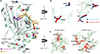 |
Figure 1 (A) The homologous PcyDHFR protein structure of wild-type (light grey), human (pale cyan), and macaque (pale green) were aligned and shown in a ribbon structure. The mutations in protein were highlighted in the navy and red spheres for human and macaque samples, respectively. (B) Superimposition of four shared mutations between human and macaque P. cynomolgi. (C) The mutations in disordered protein residues from macaque isolates are shown as red ball and stick. Dashed lines represented a two-flexible loop structure, which is not well defined in the PvDHFR template crystal structure [15], and seven amino acid insertions in a sequence of PcyDHFR protein. |
The entire PcyDHPS domain in the range of C-terminal of bifunctional protein, HPPK-DHPS at position 334th–724th was selected for 3D structural modeling based on homology with a PfDHPS template (PDB: 6JWX), which covered 90% of PcyDHPS sequences and shown sequence similarity with structure template were 66.51–66.82% for human and macaque P. cynomolgi, respectively [2]. The modeled PcyDHPS structures contained two loop structures at 411th–444th and 589th–676th, corresponding to 2 insertion sites in the PcyDHPS sequence compared to the PfDHPS sequence. The Loop-2 were combined from the insertion sequence at 589th–629th, and a not well-defined template PfDHPS structure at position 631st–676th (Fig. 2B). The human P. cynomolgi isolates carried the V451I mutation that far from the DHPS protein’s active site. In contrast, the macaque isolates presented the A585V mutation, which is equivalent to the protein binding pockets of PfDHPS and PvDHPS at residue A613 and V585, respectively, that are attributed to sulfa-drug resistance in the parasites. Hence, an in-depth analysis of the impact of the detected mutation in this region on protein affinity was conducted on the structure of the macaque isolate by protein docking and interaction analysis using the enzyme’s natural substrate (pABA) and two of its inhibitors, SDX and SMZ.
 |
Figure 2 (A) An overlay of the PcyDHPS models of 3 variants: wild-type (light grey), human (light orange), and macaque (pale cyan) isolates. The identified mutations in the study are depicted in a red ball and stick; a small box was zoomed in to view mutations in the region of loop-1. The dashed line indicates loop structures, two long insertion residues in P. cynomolgi, and a disordered PfDHPS template. The bound substrates pABA (yellow) and PtPP (green) are displayed in a ball and stick representation, while the cofactor magnesium is represented by an orange sphere. (B) Schematic protein sequence alignment of PcyDHPS against PfDHPS, which identified the two insertion sites in the PcyDHPS protein sequence (yellow) and disordered PfDHPS template structure (soft red) [2]. A dash indicates a gap in sequence alignment. |
Molecular docking between PcyDHFR and its inhibitors
We identified 24 mutations of PcyDHFR in each macaque isolate (Haplotype 2) and 4 mutations from the human isolate, which both variants carrying the C49 mutation, which is located close to the catalytic site of the protein (Fig. 1) and comparable with the C50 mutation associated with PYR resistance in P. falciparum [3, 5, 11]. However, other identified mutations were included in the structure to determine their potential consequences on protein structure and function by conducting molecular docking with their inhibitors.
The PcyDHFR was docked with three common antimalarial inhibitors, namely PYR, 1CY, and TOP, and we also docked three new antimalarials used for the treatment of resistance P. falciparum: P218, P65, and WR99210. These inhibitors were determined to bind within 6 Å of the protein’s active site, and reported in the ASP score function and normalized with its molecular weight. The resulting docking and normalized scores show a similarity between mutant and wild-type scores (Table 3). This finding suggests that these drugs were likely to retain the P. cynomolgi infection therapeutic potential.
ASP scoring function of molecular docking between PcyDHFR and PcyDHPS and its inhibitors.
Additional analysis of the highest docking score was executed by protein-ligand interaction analysis. The result revealed that the hydrogen bond interaction between the mutant protein and PYR ligand was slightly different from the wild-type. Moreover, the unique interaction at residues S123, Y185, and T200 was identified in the human P. cynomolgi (Fig. 3A). The S123 residue was equal to S108 and S117 of PfDHFR and PvDHFR, respectively, directly related to PYR resistance [15, 39]. Consequently, the differences in protein interaction were further confirmed by molecular dynamics (MD) simulation, which determined the percent hydrogen occupancy and calculated binding free energies (ΔGbind) to elucidate the interaction in detail.
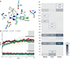 |
Figure 3 (A) Illustration H-bonding of the docked PcyDHFR-PYR complex of 3 variants P. cynomolgi. Contact residues are shown in the sticks with grey (wild-type), cyan (human), and light green (macaque). The H bonds are represented in grey, cyan, and yellow dashed lines for wild-type, human, and macaque variants, respectively. A unique hydrogen bond forming residues of the human variant is highlighted in the blue box. (B) Display RMSD analysis of the docked complex through molecular simulations. The last 20 ns were selected for further analysis, indicated in the grey area. (C) Heatmap of the hydrogen occupancy at 81–100 ns of simulation. The amino acids with unique hydrogen occupancy are shown in the square box with their occupancy percentages. |
Molecular dynamic simulation of the PcyDHFR-PYR complex
We simulated the molecular dynamics of the PcyDHFR-PYR complex of the wild-type, human, and macaque variants using GROMACS software, a widely used tool for molecular dynamics simulations. Each complex underwent three 100-ns simulations. The root mean square deviation (RMSD) plots of all variants showed a stable simulation system throughout the 100 ns of simulation (Fig. 3B). The average RMSD scores of the backbone proteins of the wild-type, human, and macaque variants were 3.58, 3.64, and 3.32 Å, respectively. In addition, the stabilizing average of the PYR ligand of these three variants was less than 1 Å with a reasonably low deviation. These values and plots indicate that the PcyDHFR-PYR complex formed a stable structure and system during the simulation. Therefore, the last 20 ns trajectories to analyze hydrogen occupancies and binding free energy.
GROMACS analysis of the three P. cynomolgi variants mostly detected one hydrogen bond interaction occurring between PcyDHFR and PYR throughout the simulation. The percent hydrogen bond occupancies of 13 wild-type residues ranged from 0.17% to 28.27%. Two of the residues had unique interactions; specifically, S126 and R137 had 2.97% and 0.35% hydrogen occupancy, respectively. Twelve residues of the human variant exhibited hydrogen occupancies ranging from 0.17% to 30.02%; moreover, positions C14, S58, and T61 (found only in this variant), showed 2.09%, 0.52%, and 0.17% hydrogen occupancies, respectively. Remarkably, no occupancies were found at the S123 and Y185 positions, and T200 carried a low occupancy of approximately 1%, consistent with the analysis of the entire simulation of 100 ns. These results suggest that these unique positions from protein interaction analysis might not be involved in the hydrogen bonding of the PcyDHFR-PYR complex in human P. cynomolgi isolates. In the macaque variant, we observed 0.02%–62.24% hydrogen occupancy across 14 residues of the protein complex. Unique occupancies were observed in I202 (0.18%) and L236 (0.04%). Most notably, the W47 and D53 positions exhibited the highest occupancies among all P. cynomolgi variants, suggesting that the formation of hydrogen bonds at these residues probably represents a crucial interaction in the PcyDHFR-PYR complex (Fig. 3C).
Binding free energies of PcyDHFR protein
The simulated complexes from the last 20 ns of the PcyDHFR molecular dynamic were collected and analyzed by MM-PBSA on GROMACS to evaluate the alteration in the binding energy of the mutant variants. Table 4 shows that the main interaction of the PcyDHFR-PYR complex was the Van der Waals interaction, with a highly negative Van der Waals energy (ΔEvdw), but we observed no significant difference between the P. cynomolgi variants (p-value > 0.05). Similarly, the other binding energies, i.e., electrostatic energy (ΔEele), polar solvation energy (ΔEpol), and solvent accessible surface area (SASA) also showed no significant difference (Table 4). The ΔGbind values of these mutants retained similar to wild-type PcyDHFR (p-value > 0.05). This result indicates that the detected missense mutations in human and macaque PcyDHFR did not influence PYR-binding affinity; therefore, the PYR drug was predicted to remain effective in treating P. cynomolgi infection.
Computational analysis of binding energies of the docked PcyDHFR-PYR complex by using MM-PBSA.
Molecular docking of PcyDHPS with a natural substrate and inhibitors
The structures of PcyDHPS were built and retrieved from SWISS-MODEL, which were established based on a homologous PfDHPS. The ligands were occupied within 3 Å of the protein binding site and reported as ASP fitness score in Table. 3. As a result of the docking score of the substrate, pABA showed a slight difference in mutant variants compared to the wild-type; similarly, the normalized scores scantily exhibited differences as well (approximate 0.01). The protein interaction analysis revealed a unique hydrogen bond interaction between human and macaque P. cynomolgi at position G551 and K581, which could be a factor in the higher score of the mutant variants.
Meanwhile, the scores of inhibitors SDX and SMZ also exhibited a trivial difference. They barely differed in the normalized score, implying that the mutations found did not alter the antimalarial binding affinity with the PcyDHPS protein. The interaction analysis of the human and macaque P. cynomolgi protein complexed with SDX showed a hydrophobic interaction with D511 and F552, which was absent in wild-type P. cynomolgi. Moreover, another hydrogen bond was detected at G551, while only one bond was present in the wild-type (Figs. 4A, 4C). In addition, a unique hydrophobic interaction was found at residue F552 and H556 in the PcyDHPS-SMZ complex of the mutant variants that was absent in the wild-type complex. All P. cynomolgi variants exhibited three hydrogen bonds with SMZ, a consequence of slightly different docking scores (Figs. 4B, 4D). These results provide insight into how the increase in docking score could be a rationale for the differential bonds in the interaction between PcyDHPS and inhibitors.
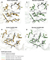 |
Figure 4 The PcyDHPS-SDX and SMZ complex interaction of human (light orange) and macaque (light green) P. cynomolgi superimposed with the wild-type interaction (grey). The dashed line represents the interaction between the protein and inhibitors, and the unique interaction residues of mutants are highlighted in yellow boxes. |
Notable, hydrogen bonds interacted with K581 and R582 present in all ligands, implying that these residues were crucial in the interaction between the ligand and PcyDHPS. Nevertheless, complementary experiments are required to investigate the interaction and the impact of mutations on sulfa drug affinity in P. cynomolgi.
Discussion
Simian malaria infections have been increasing in Southeast Asia, where long-tailed and pig-tailed macaques (Macaca nemestrina) serve as natural hosts to P. knowlesi, P. cynomolgi, and P. inui [4]. This rise is driven by factors such as deforestation, climate change, and the growing geographic overlap between macaque and human populations [14], prompting concern for public health across the region. Although P. cynomolgi infections in humans and macaques have rarely been reported in Cambodia – with one study noting a 0.96% infection rate in humans (11/14,732) and another reporting 50% prevalence in macaques (27/54) [8, 40] – multiple studies from Thailand have documented such infections more frequently [22, 23, 27]. However, few publications have explored the genotype of antifolate resistance-associated genes, including pcydhfr [13] and pcydhps, or examined the structural implications of their mutations. To address this gap, we tested all samples using two independent PCR protocols targeting the 18S rRNA [8] and cox1 genes [24], both capable of detecting a broad range of simian Plasmodium species, including P. knowlesi, P. coatneyi, P. inui, P. fieldi, and P. cynomolgi. Notably, both assays consistently detected only P. cynomolgi mono-infections, with no evidence of co-infections. Sequencing confirmed these findings, aligning with P. cynomolgi reference sequences. This could reflect regional dominance of P. cynomolgi at the time of sampling, host- or season-specific factors limiting exposure to other species, or the presence of low-level co-infections below the detection threshold.
Here, we report on four mutations in pcydhfr in humans and 24 mutations in macaques infected by P. cynomolgi. Mutations were detected based on comparisons with the reference sequence of strain M, a high-quality assembly genome that was improved and published in 2017 [20]. Loop-1 of modeled PcyDHFR differed in orientation from PfDHFR, extending from the structure [15, 39]. The S33P mutation was detected in this region. It was equal to one (P33) of six corresponding PYR resistance residues in P. vivax, which was found in Thai-Myanmar and Thai-Cambodian isolates [32]. However, the location of the S33P mutation was predicted to be far from the catalytic site of PcyDHFR and, consequently, likely has no significant role in drug resistance. Loop-2 presented a different repeat sequence in PcyDHFR from PvDHFR [15]; the relationship between the tandem repeat and protein function remains unclear. The model revealed the location of C49G in human isolates, and N44T and C49S in macaque isolates that were placed close to the binding site of PcyDHFR. The C49 was equal to the C50 of PfDHFR, one of the residues involved in controlling the active site of the protein [39], and related to a large side chain amino acids substitution [11]. Therefore, the mutant models with the mutation at C49 needed to be ascertained initially by computational analysis. The protein docking and interaction analysis of PcyDHFR with six inhibitors revealed no difference between mutants and wild-type, suggesting that the mutations do not affect protein-ligand binding. However, one unique hydrogen bond interacted with the S123 residue of human PcyDHFR-PYR complex that was equivalent to a critical residue conferring PYR and 1CY resistances in P. falciparum and P. vivax [15, 34, 39], but the simulation movement displayed no hydrogen occupancy, reflecting the hypothesis that the mechanism of this residue of the parasite was less related to direct hydrogen bond formation to the ligand. During simulation, all PcyDHFR complexes showed a single hydrogen bond formation, and a consistency in the percentage of hydrogen occupancies and contact residues was revealed. The important role of the PcyDHFR’s W47 and D53 residues in hydrogen bonding formation with its ligand was represented by the highest hydrogen occupancy in all PcyDHR-PYR complexes, which correspond to P. falciparum and P. vivax [15, 29, 38]. The contact residue of S58 in the human P. cynomolgi variant was comparable to the positions associated with PYR resistance in P. falciparum and P. vivax [9, 35]. Resulting in indirect contact with inhibitors by pointing out to the protein surface and solvent in PfDHFR, whereas PvDHFR is pointed forward into the protein active site [15, 39]. The simulation in this study elucidated the indirect impact of the S58 to PYR binding in P. cynomolgi by identifying a low percentage of hydrogen bond occupancies and unidentified hydrogen bond occupancies in this position. The VDW interaction was a major stabilizing interaction in the PcyDHFR-PYR complex. It was slightly different and favorable compared to the wild-type of PfDHFR [18]. Interestingly, the total binding energies of all complexes were not significantly different among all variants, confirming that the mutations found in PcyDHFR did not contribute to protein structure and interfere with protein-PYR affinity. However, further experiments should be carried out to confirm this finding.
This study determined that the mutation covered five equivalent residues of the DHPS binding pocket of P. falciparum and P. vivax, which confer sulfa drug resistance in the parasite. Among the detected pcydhps mutations, the A585V mutation of macaque P. cynomolgi was equal to the fifth significance residues, A613 and V585, respectively [10, 35]. In an in vitro study of PvDHPS, the V585A mutation resulted in a 2-fold increase in the Ki value of SDX, while the A383G mutation led to a 30-fold increase that is consistent with PfDHPS. This implies that the mutation at the fifth position of parasites has a low influence on the SDX binding affinity compared to the mutated residues at the first and second positions. The fifth position indirectly affects protein interaction by contributing to salt-bridge formation, which helps dimerization of the protein [2, 21, 36]. Although PcyDHPS is closely related to PvDHPS and has 90% protein sequence similarity with the available structure, the structure contains a constriction and an active site that limits the accessibility of SDX and SMZ ligands for docking in this study; accordingly, PfDHPS was selected as the template structure. Loop-1 of the build model PcyDHPS was located far from the core folding structure, and another loop contained a tandem-like repeat, like in PvDHPS, which has a function in PvHPPK-DHPS protein dimerization, providing evidence of indirect function on protein activity [36]. Molecular docking of the PcyDHPS mutant variants is only slightly different from that of the wild-type. The higher docking score of the mutant variants is likely due to a unique hydrophobic interaction involving five residues to support ligand binding and stabilize ligand alignment along the binding site of P. falciparum. All PcyDHPS variants formed a hydrogen bond with K581 and R582, which could contribute, as characterized in PfDHPS, to help the ligand to stay in the proper position and form a salt bridge to secure the substrates at the binding site [2]. The interaction analysis of PcyDHPS variants revealed the persistence of the likely crucial residue for interaction between protein and inhibitors, proposing that the detected mutations in P. cynomolgi do not alter PcyDHPS-ligand binding. This suggests the potential of sulfa drugs for treating P. cynomolgi, but further testing is required to confirm the relationship between these mutations and sulfa drug resistance in parasites.
The mutations in pcydhfr and pcydhps should be investigated further to clarify the relationship between these polymorphisms and antifolate resistance in P. cynomolgi. The haplotype pattern of the pcydhfr-pcydhps was distinct among different hosts, indicating that transmission of P. cynomolgi among hosts could be limited, or the parasite has been circulating within the host population and area. To investigate this hypothesis, an extensive sample size analysis is required to confirm a significant comparison of the results between the hosts.
The relatively high number of amino acid differences observed between the human and simian P. cynomolgi isolates may reflect natural genetic divergence rather than selective pressure from antimalarial drugs. Several of these polymorphisms were found in regions not previously associated with resistance phenotypes and may instead represent neutral mutations or lineage-specific variations. One possible explanation, particularly regarding the DHFR gene, is that a single zoonotic crossover event may have occurred in the past, followed by local expansion and sustained circulation within the human population. The human-infecting isolates may represent a genetically distinct subpopulation that has at least partially adapted to human hosts, leading to reduced gene flow with macaque parasite populations. Alternatively, these differences could indicate a cryptic transmission cycle or a founder effect, where a limited number of genetically distinct parasites crossed into humans and expanded clonally. These hypotheses need further investigation using whole-genome sequencing and broader geographic or longitudinal sampling.
In conclusion, the observed amino acid differences between human and simian P. cynomolgi isolates are more likely to reflect natural genetic divergence than antimalarial drug pressure. These variations may result from a past zoonotic crossover event, followed by local adaptation and limited gene flow with macaque populations, or may reflect a cryptic transmission cycle. Further investigation using whole-genome sequencing and expanded sampling is needed to clarify the evolutionary and epidemiological significance of these findings.
Acknowledgments
We thank Ms. Watcharee Pagornrat, Ms. Wanassanan Madmanee, Ms. Kanokon Suwannasin, and Ms. Jindarat Kouhathog for their help throughout the project.
Funding
This research project has been funded by Mahidol University (Fundamental Fund: fiscal year 2025 by National Science Research and Innovation Fund (NSRF)) to MI, and part of the Mahidol-University Oxford Tropical Medicine Research Programme funded by the Wellcome Trust of the UK (core grant 106698/B/14/Z) and Wellcome OA statement. This research was funded in whole, or in part, by the Wellcome Trust [220211]. For the purpose of Open Access, the author has applied a CC BY public copyright license to any Author Accepted Manuscript version arising from this submission. The funding sources did not participate in data analysis or the final decision to publish the manuscript. This project is funded by National Research Council of Thailand (NRCT): Contract number: N42A680526, the Research Fund Senior Scholar (grant no. RTA6280010, to S.M.).
Conflicts of interest
The authors declare that they have no competing interests.
Data availability statement
All data generated or analyzed during this study are included in this published article.
Author contributions statement
RS, AMD, and MI contributed to study design. AMD and NPJD acquired funding for the study. SM collected all samples and data. RS prepared, genotyped, and sequenced DNA. NJ performed the simulation. RS, KC, and MI analyzed the data. RS and MI wrote the report. All authors read and approved the final manuscript.
Supplementary material
Supplementary File 1: Nucleotide sequences of pcydhfr.
Supplementary File 2: Nucleotide sequences of pcydhps.
Access Supplementary MaterialReferences
- BIOVIA. 2020. Discovery Studio Visualizer. Available at https://www.3ds.com/products/biovia/discovery-studio/visualization. [Google Scholar]
- Chitnumsub P, Jaruwat A, Talawanich Y, Noytanom K, Liwnaree B, Poen S, Yuthavong Y. 2020. The structure of Plasmodium falciparum hydroxymethyldihydropterin pyrophosphokinase-dihydropteroate synthase reveals the basis of sulfa resistance. FEBS Journal, 287(15), 3273–3297. [Google Scholar]
- Chusacultanachai S, Thiensathit P, Tarnchompoo B, Sirawaraporn W, Yuthavong Y. 2002. Novel antifolate resistant mutations of Plasmodium falciparum dihydrofolate reductase selected in Escherichia coli. Molecular and Biochemical Parasitology, 120(1), 61–72. [Google Scholar]
- Coatney GR, Collins WE, Warren M, Contacos PG. 1971. The primate malarias. US National Institute of Allergy and Infectious Diseases. Rockville. Bethesda: US Department of Health, Education, and Welfare, Public Health Service, National Institutes of Health. [Google Scholar]
- Contreras CE, Cortese JF, Caraballo A, Plowe CV. 2002. Genetics of drug-resistant Plasmodium falciparum malaria in the Venezuelan state of Bolivar, American Journal of Tropical Medicine and Hygiene, 67(4), 400–405. [Google Scholar]
- Genheden S, Ryde U. 2015. The MM/PBSA and MM/GBSA methods to estimate ligand-binding affinities. Expert Opinion on Drug Discovery, 10(5), 449–461. [Google Scholar]
- Hyde JE. 1990. The dihydrofolate reductase-thymidylate synthetase gene in the drug resistance of malaria parasites. Pharmacology and Therapeutics, 48(1), 45–59. [Google Scholar]
- Imwong M, Madmanee W, Suwannasin K, Kunasol C, Peto TJ, Tripura R, von Seidlein L, Nguon C, Davoeung C, Day NPJ, Dondorp AM, White NJ. 2019. Asymptomatic natural human infections with the simian malaria parasites Plasmodium cynomolgi and Plasmodium knowlesi. Journal of Infectious Diseases, 219(5), 695–702. [CrossRef] [PubMed] [Google Scholar]
- Imwong M, Pukrittakayamee S, Looareesuwan S, Pasvol G, Poirreiz J, White NJ, Snounou G. 2001. Association of genetic mutations in Plasmodium vivax dhfr with resistance to sulfadoxine-pyrimethamine: geographical and clinical correlates. Antimicrobial Agents and Chemotherapy, 45(11), 3122–3127. [Google Scholar]
- Imwong M, Pukrittayakamee S, Cheng Q, Moore C, Looareesuwan S, Snounou G, White NJ, Day NP. 2005. Limited polymorphism in the dihydropteroate synthetase gene (dhps) of Plasmodium vivax isolates from Thailand. Antimicrobial Agents and Chemotherapy, 49(10), 4393–4395. [Google Scholar]
- Japrung D, Leartsakulpanich U, Chusacultanachai S, Yuthavong Y. 2007. Conflicting requirements of Plasmodium falciparum dihydrofolate reductase mutations conferring resistance to pyrimethamine-WR99210 combination. Antimicrobial Agents and Chemotherapy, 51(12), 4356–4360. [Google Scholar]
- Jones G, Willett P, Glen RC, Leach AR, Taylor R. 1997. Development and validation of a genetic algorithm for flexible docking. Journal of Molecular Biology, 267(3), 727–748. [Google Scholar]
- Karnchaisri K, Day NPJ, Dondorp AM, Malaivijitnond S, Imwong M. 2024. Prevalence and genetic diversity of simian malaria in wild macaque populations across Thailand: Implications for human health. Acta Tropica, 254, 107187. [Google Scholar]
- Kojom Foko LP, Kumar A, Hawadak J, Singh V. 2023. Plasmodium cynomolgi in humans: current knowledge and future directions of an emerging zoonotic malaria parasite. Infection, 51(3), 623–640. [Google Scholar]
- Kongsaeree P, Khongsuk P, Leartsakulpanich U, Chitnumsub P, Tarnchompoo B, Walkinshaw MD, Yuthavong Y. 2005. Crystal structure of dihydrofolate reductase from Plasmodium vivax: pyrimethamine displacement linked with mutation-induced resistance. Proceedings of the National Academy of Sciences of the United States of America, 102(37), 13046–13051. [Google Scholar]
- Lee KS, Divis PC, Zakaria SK, Matusop A, Julin RA, Conway DJ, Cox-Singh J, Singh B. 2011. Plasmodium knowlesi: reservoir hosts and tracking the emergence in humans and macaques, PLOS Pathogens, 7(4), e1002015. [Google Scholar]
- Malde AK, Zuo L, Breeze M, Stroet M, Poger D, Nair PC, Oostenbrink C, Mark AE. 2011. An automated force field topology builder (ATB) and repository: version 1.0. Journal of chemical theory and computation, 7(12), 4026–4037. [Google Scholar]
- Mokmak W, Chunsrivirot S, Hannongbua S, Yuthavong Y, Tongsima S, Kamchonwongpaisan S. 2014. Molecular dynamics of interactions between rigid and flexible antifolates and dihydrofolate reductase from pyrimethamine-sensitive and pyrimethamine-resistant Plasmodium falciparum. Chemical Biology & Drug Design, 84(4), 450–461. [Google Scholar]
- Muehlenbein MP, Ancrenaz M, Sakong R, Ambu L, Prall S, Fuller G, Raghanti MA. 2012. Ape conservation physiology: fecal glucocorticoid responses in wild Pongo pygmaeus morio following human visitation. PLoS One, 7(3), e33357. [Google Scholar]
- Pasini EM, Bohme U, Rutledge GG, Voorberg-Van der Wel A, Sanders M, Berriman M, Kocken CH, Otto TD. 2017. An improved Plasmodium cynomolgi genome assembly reveals an unexpected methyltransferase gene expansion. Wellcome Open Research, 2, 42. [Google Scholar]
- Pornthanakasem W, Riangrungroj P, Chitnumsub P, Ittarat W, Kongkasuriyachai D, Uthaipibull C, Yuthavong Y, Leartsakulpanich U. 2016. Role of Plasmodium vivax dihydropteroate synthase polymorphisms in sulfa drug resistance. Antimicrobial Agents and Chemotherapy, 60(8), 4453–4463. [Google Scholar]
- Putaporntip C, Jongwutiwes S, Thongaree S, Seethamchai S, Grynberg P, Hughes AL. 2010. Ecology of malaria parasites infecting Southeast Asian macaques: evidence from cytochrome b sequences. Molecular Ecology, 19(16), 3466–3476. [CrossRef] [PubMed] [Google Scholar]
- Putaporntip C, Kuamsab N, Pattanawong U, Yanmanee S, Seethamchai S, Jongwutiwes S. 2021. Plasmodium cynomolgi co-infections among symptomatic malaria patients, Thailand. Emerging Infectious Diseases, 27(2), 590–593. [Google Scholar]
- Putaporntip C, Kuamsab N, Seethamchai S, Pattanawong U, Rojrung R, Yanmanee S, Cheng CW, Jongwutiwes S. 2021. Cryptic Plasmodium inui and P. fieldi infections among symptomatic malaria patients in Thailand. Clinical Infectious Diseases, 75(5): 805–812. [Google Scholar]
- R Core Team. 2023. R: A language and environment for statistical computing. Available at https://www.R-project.org/. [Google Scholar]
- Roth A, Maher SP, Conway AJ, Ubalee R, Chaumeau V, Andolina C, Kaba SA, Vantaux A, Bakowski MA, Thomson-Luque R, Adapa SR, Singh N, Barnes SJ, Cooper CA, Rouillier M, McNamara CW, Mikolajczak SA, Sather N, Witkowski B, Campo B, Kappe SHI, Lanar DE, Nosten F, Davidson S, Jiang RHY, Kyle DE, Adams JH. 2018. A comprehensive model for assessment of liver stage therapies targeting Plasmodium vivax and Plasmodium falciparum. Nature Communications, 9(1), 1837. [Google Scholar]
- Sai-Ngam P, Pidtana K, Suida P, Poramathikul K, Lertsethtakarn P, Kuntawunginn W, Tadsaichol S, Arsanok M, Sornsakrin S, Chaisatit C, Mathavarat C, Thaloengsok S, Boonyarangka P, Thongpiam C, Demons S, Vesely B, Waters NC, Saejeng A, Wojnarski M, Tabprasit S, Kwanpichit C, Griesenbeck JS, Spring M. 2022. Case series of three malaria patients from Thailand infected with the simian parasite, Plasmodium cynomolgi. Malaria Journal, 21(1), 142. [Google Scholar]
- Schrödinger L. 2021. The PyMOL molecular graphics system. [Google Scholar]
- Sirawaraporn W, Sirawaraporn R, Yongkiettrakul S, Anuwatwora A, Rastelli G, Kamchonwongpaisan S, Yuthavong Y. 2002. Mutational analysis of Plasmodium falciparum dihydrofolate reductase: the role of aspartate 54 and phenylalanine 223 on catalytic activity and antifolate binding. Molecular and Biochemical Parasitology, 121(2), 185–193. [Google Scholar]
- Swanson JM, Henchman RH, McCammon JA. 2004. Revisiting free energy calculations: a theoretical connection to MM/PBSA and direct calculation of the association free energy. Biophysical Journal, 86(1 Pt 1): 67–74. [Google Scholar]
- Tamura K, Stecher G, Kumar S. 2021. MEGA11: molecular evolutionary genetics analysis version 11. Molecular Biology and Evolution, 38(7), 3022–3027. [CrossRef] [PubMed] [Google Scholar]
- Thongdee P, Kuesap J, Rungsihirunrat K, Tippawangkosol P, Mungthin M, Na-Bangchang K. 2013. Distribution of dihydrofolate reductase (dhfr) and dihydropteroate synthase (dhps) mutant alleles in Plasmodium vivax isolates from Thailand. Acta Tropica, 128(1), 137–143. [Google Scholar]
- Triglia T, Cowman AF. 1994. Primary structure and expression of the dihydropteroate synthetase gene of Plasmodium falciparum. Proceedings of the National Academy of Sciences of the United States of America, 91(15), 7149–7153. [Google Scholar]
- Vanichtanankul J, Taweechai S, Uttamapinant C, Chitnumsub P, Vilaivan T, Yuthavong Y, Kamchonwongpaisan S. 2012. Combined spatial limitation around residues 16 and 108 of Plasmodium falciparum dihydrofolate reductase explains resistance to cycloguanil. Antimicrobial Agents and Chemotherapy, 56(7), 3928–3935. [Google Scholar]
- Wang P, Lee CS, Bayoumi R, Djimde A, Doumbo O, Swedberg G, Dao LD, Mshinda H, Tanner M, Watkins WM, Sims PF, Hyde JE. 1997. Resistance to antifolates in Plasmodium falciparum monitored by sequence analysis of dihydropteroate synthetase and dihydrofolate reductase alleles in a large number of field samples of diverse origins. Molecular and Biochemical Parasitology, 89(2), 161–177. [Google Scholar]
- Yogavel M, Nettleship JE, Sharma A, Harlos K, Jamwal A, Chaturvedi R, Sharma M, Jain V, Chhibber-Goel J, Sharma A. 2018. Structure of 6-hydroxymethyl-7, 8-dihydropterin pyrophosphokinase-dihydropteroate synthase from Plasmodium vivax sheds light on drug resistance. Journal of Biological Chemistry, 293(39), 14962–14972. [Google Scholar]
- Yusuf NM, Zulkefli J, Jiram AI, Vythilingam I, Hisam S, Devi R, Salehhuddin A, Ali NM, Isa M, Alias N, Ogu Salim N, Aziz AA, Sulaiman LH. 2022. Plasmodium spp. in macaques, Macaca fascicularis, in Malaysia, and their potential role in zoonotic malaria transmission. Parasite, 29, 32. [Google Scholar]
- Yuthavong Y, Yuvaniyama J, Chitnumsub P, Vanichtanankul J, Chusacultanachai S, Tarnchompoo B, Vilaivan T, Kamchonwongpaisan S. 2005. Malarial (Plasmodium falciparum) dihydrofolate reductase-thymidylate synthase: structural basis for antifolate resistance and development of effective inhibitors. Parasitology, 130(Pt 3), 249–259. [Google Scholar]
- Yuvaniyama J, Chitnumsub P, Kamchonwongpaisan S, Vanichtanankul J, Sirawaraporn W, Taylor P, Walkinshaw MD, Yuthavong Y. 2003. Insights into antifolate resistance from malarial DHFR-TS structures. Nature Structural Biology, 10(5), 357–365. [Google Scholar]
- Zhang X, Kadir KA, Quintanilla-Zarinan LF, Villano J, Houghton P, Du H, Singh B, Smith DG. 2016. Distribution and prevalence of malaria parasites among long-tailed macaques (Macaca fascicularis) in regional populations across Southeast Asia. Malaria Journal, 15(1), 450. [Google Scholar]
Cite this article as: Sangsri R, Jongkon N, Choowongkomon K, Malaivijitnond S, Day NPJ, Dondorp AM & Imwong M. 2025. Molecular analysis of DHFR and DHPS gene mutations in Plasmodium cynomolgi from humans and macaques in Southeast Asia. Parasite 32, 64. https://doi.org/10.1051/parasite/2025057.
All Tables
Comparison analysis of polymorphic alleles PcyDHFR and PcyDHPS. The bold blue text indicates different residues among three strains of P. cynomolgi. Bold text represents the polymorphic alleles PcyDHFR and PcyDHPS in this study.
ASP scoring function of molecular docking between PcyDHFR and PcyDHPS and its inhibitors.
Computational analysis of binding energies of the docked PcyDHFR-PYR complex by using MM-PBSA.
All Figures
|
Figure 1 (A) The homologous PcyDHFR protein structure of wild-type (light grey), human (pale cyan), and macaque (pale green) were aligned and shown in a ribbon structure. The mutations in protein were highlighted in the navy and red spheres for human and macaque samples, respectively. (B) Superimposition of four shared mutations between human and macaque P. cynomolgi. (C) The mutations in disordered protein residues from macaque isolates are shown as red ball and stick. Dashed lines represented a two-flexible loop structure, which is not well defined in the PvDHFR template crystal structure [15], and seven amino acid insertions in a sequence of PcyDHFR protein. |
| In the text | |
|
Figure 2 (A) An overlay of the PcyDHPS models of 3 variants: wild-type (light grey), human (light orange), and macaque (pale cyan) isolates. The identified mutations in the study are depicted in a red ball and stick; a small box was zoomed in to view mutations in the region of loop-1. The dashed line indicates loop structures, two long insertion residues in P. cynomolgi, and a disordered PfDHPS template. The bound substrates pABA (yellow) and PtPP (green) are displayed in a ball and stick representation, while the cofactor magnesium is represented by an orange sphere. (B) Schematic protein sequence alignment of PcyDHPS against PfDHPS, which identified the two insertion sites in the PcyDHPS protein sequence (yellow) and disordered PfDHPS template structure (soft red) [2]. A dash indicates a gap in sequence alignment. |
| In the text | |
|
Figure 3 (A) Illustration H-bonding of the docked PcyDHFR-PYR complex of 3 variants P. cynomolgi. Contact residues are shown in the sticks with grey (wild-type), cyan (human), and light green (macaque). The H bonds are represented in grey, cyan, and yellow dashed lines for wild-type, human, and macaque variants, respectively. A unique hydrogen bond forming residues of the human variant is highlighted in the blue box. (B) Display RMSD analysis of the docked complex through molecular simulations. The last 20 ns were selected for further analysis, indicated in the grey area. (C) Heatmap of the hydrogen occupancy at 81–100 ns of simulation. The amino acids with unique hydrogen occupancy are shown in the square box with their occupancy percentages. |
| In the text | |
|
Figure 4 The PcyDHPS-SDX and SMZ complex interaction of human (light orange) and macaque (light green) P. cynomolgi superimposed with the wild-type interaction (grey). The dashed line represents the interaction between the protein and inhibitors, and the unique interaction residues of mutants are highlighted in yellow boxes. |
| In the text | |
Current usage metrics show cumulative count of Article Views (full-text article views including HTML views, PDF and ePub downloads, according to the available data) and Abstracts Views on Vision4Press platform.
Data correspond to usage on the plateform after 2015. The current usage metrics is available 48-96 hours after online publication and is updated daily on week days.
Initial download of the metrics may take a while.