Figure 3
Download original image
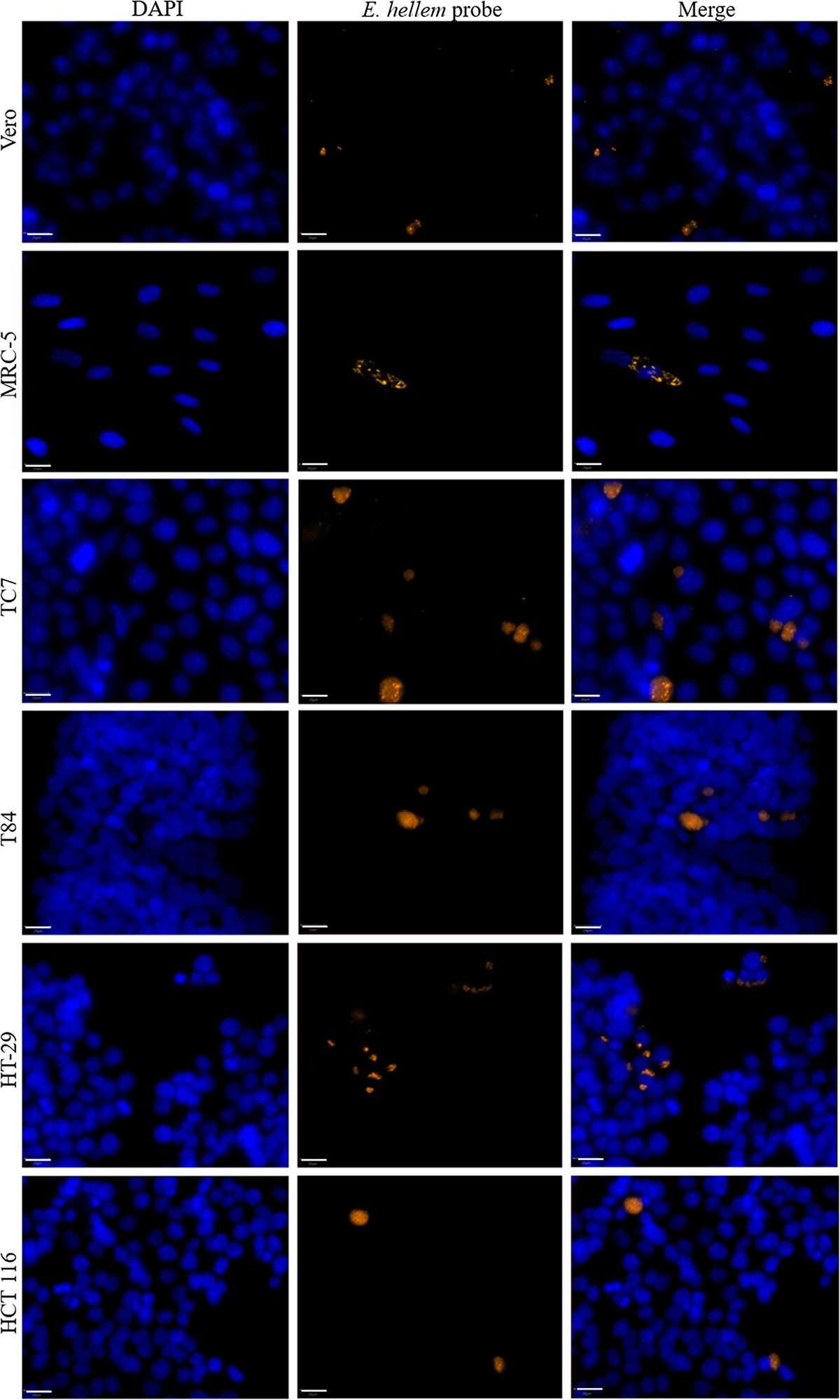
Surface areas of three species of Encephalitozoon in six cell lines. Measurement of the surface area of infectious foci within cells infected by E. intestinalis, E. hellem, and E. cuniculi at 48 h post-infection. The results were obtained from three biological replicates. The number of areas measured is indicated under each cell line. The median is represented by a solid line. The overall p-value is shown in parentheses for each species. Post hoc pairwise comparisons are indicated by asterisks: *p ≤ 0.05, **p ≤ 0.01, ***p ≤ 0.001.
Current usage metrics show cumulative count of Article Views (full-text article views including HTML views, PDF and ePub downloads, according to the available data) and Abstracts Views on Vision4Press platform.
Data correspond to usage on the plateform after 2015. The current usage metrics is available 48-96 hours after online publication and is updated daily on week days.
Initial download of the metrics may take a while.