| Issue |
Parasite
Volume 33, 2026
|
|
|---|---|---|
| Article Number | 17 | |
| Number of page(s) | 8 | |
| DOI | https://doi.org/10.1051/parasite/2026017 | |
| Published online | 01 April 2026 | |
Research Article
A novel RFLP-PCR method for the rapid diagnosis of Echinococcus multilocularis and different Echinococcus granulosus sensu lato species from tissue samples
Une nouvelle méthode RFLP-PCR pour le diagnostic rapide d’Echinococcus multilocularis et de différentes espèces d’Echinococcus granulosus sensu lato à partir d’échantillons tissulaires
1
INTERFAS Unit, ANSES, National Reference Laboratory for Echinococcus spp., Rabies and Wildlife Laboratory, 54220 Malzéville, France
2
UMR BIPAR, ANSES, Laboratoire de Santé Animale, INRAE, École Nationale Vétérinaire d’Alfort, 94700 Maisons-Alfort, France
* Corresponding authors: This email address is being protected from spambots. You need JavaScript enabled to view it.
; This email address is being protected from spambots. You need JavaScript enabled to view it.
Received:
30
September
2024
Accepted:
26
February
2026
Abstract
Both alveolar echinococcosis and cystic echinococcosis are zoonotic diseases affecting thousands of humans in Europe annually. Five Echinococcus species are known to be endemic in Europe, but with varying relative importance in humans and animals. In order to identify these Echinococcus species from tissue samples, an RFLP-PCR method was developed and validated in accordance with the French quality standards for molecular diagnostics (NF U47-600-2). Amplification by PCR of about 846 bp from the 12S ribosomal RNA gene was followed by a single digestion step with three restriction enzymes (AccI, DdeI and HinfI). The RFLP-PCR method distinguishes Echinococcus multilocularis, E. granulosus sensu stricto, E. equinus, E. ortleppi and E. canadensis in six specific profiles composed of two or three bands. Each profile is specific for the species, except an additional specific profile from genotype G8 of E. canadensis. Species from other genera of the Taeniidae family correspond to two other types of profiles. Additionally, in silico analyses predicted three specific additional profiles for the other four Echinococcus species. The limit of detection of the method including DNA extraction is estimated to be a minimum of 1,000 copies. Total specificity and sensitivity were obtained by testing a panel of 73 and 79 DNA samples previously identified by sequencing. This RFLP-PCR technique is cost-effective, simple and rapid to implement, making it suitable for use in large epidemiological studies, as well as for single diagnostic purposes targeting tissue samples, such as cysts or worms, without requiring sequencing.
Résumé
L’échinococcose alvéolaire et l’échinococcose kystique sont des zoonoses qui affectent chaque année des milliers de personnes en Europe. Cinq espèces d’Echinococcus sont endémiques en Europe, mais leur importance relative chez l’homme et l’animal varie. Afin d’identifier ces espèces d’Echinococcus à partir d’échantillons tissulaires, une méthode RFLP-PCR a été développée et validée conformément aux normes françaises de qualité pour le diagnostic moléculaire (NF U47-600-2). L’amplification par PCR d’un fragment d’environ 846 pb du gène de l’ARN ribosomique 12S est suivie d’une digestion unique par trois enzymes de restriction (AccI, DdeI et HinfI). La méthode RFLP-PCR permet de distinguer Echinococcus multilocularis, E. granulosus sensu stricto, E. equinus, E. ortleppi et E. canadensis grâce à six profils spécifiques composés de deux ou trois bandes. Chaque profil est spécifique de l’espèce, à l’exception d’un profil spécifique supplémentaire associé au génotype G8 d’E. canadensis. Les espèces appartenant à d’autres genres de la famille des Taeniidae correspondent à deux autres types de profils. De plus, des analyses bioinformatiques ont prédit trois profils spécifiques supplémentaires pour les quatre autres espèces d’Echinococcus. La limite de détection de la méthode, extraction d’ADN incluse, est estimée à au moins 1 000 copies. La spécificité et la sensibilité totales ont été obtenues en testant un panel de 73 et 79 échantillons d’ADN préalablement identifiés par séquençage. Le faible coût, la simplicité et la rapidité de mise en œuvre de cette technique RFLP-PCR la rendent adaptée aux études épidémiologiques de grande envergure, ainsi qu’au diagnostic ciblé d’échantillons tissulaires, tels que des kystes ou des vers, sans nécessiter de séquençage.
Key words: Echinococcus / Molecular diagnostic / RFLP-PCR / Larval stage
Edited by: Frédéric Grenouillet
Equal contribution
© G. Umhang et al., published by EDP Sciences, 2026
 This is an Open Access article distributed under the terms of the Creative Commons Attribution License (https://creativecommons.org/licenses/by/4.0), which permits unrestricted use, distribution, and reproduction in any medium, provided the original work is properly cited.
Introduction
The genus Echinococcus, which belongs to the family Taeniidae, is currently composed of nine different species [20, 41]. The most common species are Echinococcus multilocularis and those of Echinococcus granulosus sensu lato (s.l.). The latter is currently recognised as a complex of five species: Echinococcus granulosus sensu stricto (s.s.), E. equinus, E. ortleppi, E. canadensis and E. felidis. The taxonomy of E. canadensis remains unresolved, with four genotypes (G6, G7, G8 and G10) grouped together, which may be divided into two or three distinct species [18, 26, 29]. With the exception of E. felidis, which is exclusively distributed in Africa, all other species of E. granulosus s.l. are present in Europe as E. multilocularis and have been shown to be zoonotic, but with varying relative importance.
Echinococcus multilocularis and E. granulosus s.l. are the etiological agent of alveolar echinococcosis (AE) and cystic echinococcosis (CE), respectively. Both are chronic diseases with initial asymptomatic periods that can last up to 15 years. Compared to CE, which is a disabling disease with a low mortality rate, AE is more severe and has a high mortality rate without treatment [4]. The average annual incidence of AE in humans in the 27 Member States of the European Union was 151 cases per year between 2018 and 2022 [7]. From 1997 to 2021, 54,244 cases of human CE were identified in the 27 Member States [5]. The distinction between AE and CE in humans is typically made on the basis of imaging and serological techniques rather than molecular diagnostics, which is not always possible, and which is not consistently reported in official statistics. Furthermore, the identification of the E. granulosus s.l. species involved in CE cases, which necessarily requires molecular biology, is even less systematic. In this context, a systematic review of 599 molecularly confirmed human CE cases from 29 European countries between 2000 and 2021 showed that E. granulosus s.s. was the most common species (76.8%), followed by the E. canadensis cluster (21.7%) and E. ortleppi (1.2%) [6]. The recent identification of the first human case due to E. equinus has raised questions about its potential for transmission to humans [22].
In Europe, E. multilocularis is maintained by a sylvatic lifecycle based on the predation of voles (Microtus arvalis and Arvicola spp.) by red foxes. A significant number of mammals, acting as either intermediate or dead-end hosts, have been identified as having the potential to develop AE. These include aquatic rodents, pigs, wild boars, horses, and a range of other exotic species commonly found in zoos or wildlife parks, such as lemurs and monkeys [25]. The European species of E. granulosus s.l. mainly have domestic life cycles involving dogs and livestock. Although other canid species, particularly wolves, can act as definitive hosts, there is typically a predominance of one type of intermediate host species for each E. granulosus species. Echinococcus granulosus s.s. is of primary concern for sheep, but it may also infect a wide range of other intermediate hosts in livestock, including cattle and pigs. In addition, it can infect wildlife species, including wild boar and cervids. Additionally, E. equinus and E. ortleppi are more specific and are primarily associated with equids (horses and donkeys) and cattle, respectively. Genotype G7 of E. canadensis is primarily associated with pigs and wild boar in Europe, while G6 concerns camels in North Africa, the Near and Middle East, and Central Asia. In contrast, genotypes G8 and G10, which are present only in northern Eurasia, have been observed to infect wild cervid species (moose, elk/wapiti and roe deer) as intermediate hosts.
The progress made in the field of molecular biology over the past four decades has been pivotal in the diagnosis of Echinococcus species from tissue samples. The morphological identification of worms has typically enabled differentiation between E. multilocularis and E. granulosus s.l. However, distinguishing between the various species within the E. granulosus complex has remained challenging, particularly given that some variations may be attributed to the definitive host species and the quality of the samples, which can often be a limitation when dealing with wildlife samples. Furthermore, the diagnostic process at the species level proved particularly challenging with regard to the larval stage. It was not possible to distinguish between species in intermediate or accidental hosts, particularly within the E. granulosus complex, either in animal or human samples. Molecular methods are currently regarded as the most pertinent approaches, with polymerase chain reaction (PCR) techniques targeting a single species or multiplex PCR representing the most effective techniques. Nevertheless, sequencing is typically conducted on the amplicons obtained from the initial PCR to confirm the species or to identify the species targeted by the PCR. This is exemplified by the commonly used Cest Multiplex from Trachsel et al. [30] in order to identify the E. granulosus species. One of the most common techniques is the use of PCR to amplify a fragment of the mitochondrial genome (e.g., cox1, nad1 and nad5) using primers efficient for a wide range of parasite species. The required sequencing to identify the Echinococcus species in question increases the time needed for analysis, as well as the associated costs. The cost, in turn, presents a barrier to use of the technique in low-income countries, yet it is often required for more accurate characterization of the lifecycles involved. The restriction fragment length polymorphism-PCR (RFLP-PCR) method offers several advantages, including rapidity, simplicity and cost-effectiveness due to the generation of a wide range of specific band patterns following electrophoresis.
Since the 1990 s, a number of RFLP-PCR assays have been developed for use with Echinococcus species. The primary differentiating factor between these tests is the target species, which corresponds to disparate epidemiologic scenarios. For example, the ability to distinguish between E. multilocularis, E. granulosus s.s. and E. shiquicus was particularly important for understanding the epidemiology of echinococcosis on the Tibetan plateau of China [43]. Similarly, the approach developed by Hüttner et al. [11] enabled the identification of E. felidis among other E. granulosus species from taeniid eggs isolated in African wild carnivores and cysts of livestock species. Globally, RFLP-PCR assays have focused primarily on distinguishing between E. granulosus species, with E. multilocularis often omitted, even in the context of endemic European populations [9, 27, 28]. This article presents a novel RFLP-PCR diagnostic method targeting all five endemic European Echinococcus species. The methodology has been validated in accordance with the French quality standards for molecular diagnostics (NF 47-600-2).
Materials and methods
Parasitic material
A genomic DNA reference sample from E. multilocularis was used for the purpose of evaluating the methodology. This reference DNA was obtained from the metacestode stage of a parasite strain, which was maintained experimentally on Swiss EOPS mice (APAFIS #34612-2022011114104529). The DNA from this parasitic tissue was extracted manually using a standard tissue kit (QIAGEN, Hilden, Germany). The number of mitochondrial genome copies was determined using a specific real-time PCR for E. multilocularis [16], as described by Umhang et al. [32]. To evaluate method sensitivity and specificity, DNA from field samples of different E. multilocularis and E. granulosus s.l. species, as well as from other genera within the Taeniidae family (Taenia, Hydatigera and Versteria), which were already available at the laboratory, were used. Prior to the present study, all samples underwent species identification through short cox1 sequencing, as described by Bowles et al. [3].
RFLP-PCR method
In the context of method evaluation, DNA extraction was performed using a commercially available kit (Maxwell RSC Blood DNA Kit) with the Maxwell48 automatic extractor (Promega, Madison, WI, USA) based on purification with magnetic particles. A piece of tissue weighing approximately 10–20 mg was initially digested with proteinase K. Following centrifugation, the supernatant was transferred to the first well of the cartridge, which was then placed in the instrument for the subsequent washing steps, concluding with elution in 100 μL.
The two PCR primers Taen F (5′–GTTTGCCACCTCGATGTTGACT–3′) and ITMTn R (5′–CTCAATAATAATCGAGGGTGACGG–3′) used in this method were initially published by Geysen et al. 2007 [8] and reported to be specific to the Taenia genus in the context of identification of Taenia saginata in muscle lesions by RFLP-PCR. Nevertheless, while amplification of about 846 bp from the 12S ribosomal RNA is obtained for species of the Taenia genus, it is also possible for other species of the Taeniidae family, including the Echinococcus genus. The reaction mixture consisting of 5 μL of 10× PCR buffer (Invitrogen, Carlsbad, CA, USA), 1.5 μL of 50 mM MgCl2 solution, 1 μL of 10 mM dNTP mix, 2.5 μL of each primer (Taen F and ITMTn R, 10 μM each), 0.2 μL of Platinum Taq DNA polymerase, 3 μL of DNA template and enough nuclease-free water was prepared to reach 50 μL of final volume for each reaction. The protocol for PCR amplification was initiated with a denaturation step at 94 °C (2 min), followed by repetition of 35 cycles composed of denaturation at 94 °C (45 s), annealing at 57 °C (45 s) and extension at 72 °C (60 s), ending with final extension at 72 °C (7 min). The PCR products were separated on a 1.5% agarose gel stained with SYBR Safe DNA Gel Stain (Invitrogen). A negative control (3 μL of nuclease-free water) was run in agarose gel with the RFLP products and positive controls composed of 3 μL of suspected Echinococcus species DNA. After confirmation of the presence a PCR product, 6 μL of the PCR product of each sample were incubated for 3 h at 37 °C with 9 μL of a mixture containing AccI, DdeI and HinfI restriction enzymes (corresponding to 2.7 units for each of the three) and 1.3 μL of 10× rCutSmart™ Buffer (New England Biolabs, Ipswich, MA, USA) in 7.5 μL of nuclease-free water. The restriction fragments were separated by electrophoresis in 3% MetaPhor agarose gel (Lonza) for 45 min at 110 V for medium size (15 cm). A DNA size marker of 100 bp was included for size identification of the bands with Uvitec 1D software (Uvitec Ltd., Cambridge, UK). In the absence of amplification, the DNA extracts were diluted (1/10) and reamplified.
Validation of the RFLP-PCR method
Limit of detection
The limit of detection (LOD) of the PCR is the smallest number of copies of target nucleic acid per unit volume that can be detected in 95% of cases. To determine this limit, three independent ranges of six different dilutions were used. For each range point, 8 replicates were performed, yielding 24 results per assay point tested. The approximate value of the LOD corresponds to the dilution giving at least 23 positive results out of 24. The LOD of the full method (DNA extraction followed by RFLP-PCR) corresponds to the minimum quantity of biological target that must initially be present in the sample in order to obtain an interpretable profile. The test sample matrix corresponds to 20 mg liver tissue sample from a naïve Swiss EOPS mouse spiked with a different number of copies of the E. multilocularis reference DNA. Each extracted DNA was tested 8 times in two independent sessions for each of the dilutions tested. The LOD was estimated as the last dilution level where all 8 replicates are positive.
Specificity and sensitivity evaluation
Diagnostic specificity was estimated from 73 tissue samples corresponding to suspicion of E. multilocularis or E. granulosus sensu lato which was diagnosed as negative (n = 32: no amplification by short cox1 PCR or non-parasitic result after sequencing) or corresponding to infection by Taeniidae species other than from the Echinococcus genus (n = 41: Taenia hydatigena, T. krabbei, T. polyacantha, Hydatigera kamiyai and Versteria mustelae) due to short cox1 sequence analysis (Table 1). Diagnostic sensitivity was estimated from 79 positive tissue samples from the laboratory collection for which the presence of E. multilocularis or an E. granulosus s.l. species (E. granulosus sensu stricto, E. equinus, E. ortleppi and E. canadensis) had previously been confirmed. Regarding E. multilocularis, samples representing the four different clades (European, Asian, North American and Mongolian) were tested as the different genotypes of E. granulosus s.l. (Table 2). Regarding Echinococcus species not available at the laboratory (E. shiquicus, E. vogeli and E. oligarthra), an in silico analysis was carried out using Geneious prime software to obtain the expected profile bands.
List of samples according to parasite and host species used to evaluate the specificity of the PCR-RFLP assay. The asterisk indicates worms as the matrix, while all the other samples concern the larval stage.
List of samples corresponding to the five Echinococcus species targeted by the PCR-RFLP assay in the context of sensitivity evaluation. The asterisk indicates worms as the matrix, while all the other samples concern the larval stage.
RFLP banding patterns for all the Echinococcus species (genotypes concerned indicated in parentheses) and other taeniid species tested obtained after digestion by DdeI, HinfI and AccI restriction enzymes of the 12S ribosomal RNA amplicon. The various bands are distinguished from one another by a slash, whereas fragments that are not identical but of comparable size, which subsequently form a single band following electrophoresis, are grouped together in brackets. For species marked with an asterisk, the in silico band profile could not be validated by a biological test.
Method robustness
The robustness of the method was evaluated by testing variations of different conditions in the amplification of E. multilocularis reference DNA. A variation in annealing temperature from 61 to 71 °C by increments of 2 °C was tested with quantities of DNA ranging from 0.1 to 10 ng. Concentrations of primers from 0.25, 0.5, 1 and 1.5 mM and from 0.5, 1, 1.5 and 2 mM of magnesium were tested with 0.1 ng of E. multilocularis DNA. The potential impact of variation in the enzymatic digestion temperature was evaluated from 33 to 41 °C for 0.1 to 10 ng of E. multilocularis reference DNA.
Results
Description of the Echinococcus specific profiles
The RFLP-PCR method distinguishes E. multilocularis, E. granulosus s.s., E. equinus, E. ortleppi and E. canadensis in six specific profiles composed of two or three bands (Fig. 1 and Table 1). Each profile is specific for the species, except the profile from genotype G8 of E. canadensis which was different from the one obtained for the other genotypes (G6, G7 and G10) of this species. Based on in silico analyses, the profile of genotype G10 is slightly different (3 bp) from genotypes G6 and G7 but appears identical after electrophoresis. Species from other genera of the Taeniidae family correspond to two other types of profiles composed of two bands. Additionally, in silico analyses predicted three specific additional profiles for the four other Echinococcus species. The same profile is expected for E. vogeli and E. oligarthra, while two others are obtained for E. felidis and E. shiquicus. Globally, 9 different profiles were obtained for the 9 Echinococcus species (including G1 and G3 genotypes for E. granulosus s.s. and the four genotypes of E. canadensis) and two others for the other Taeniidae species.
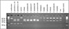 |
Figure 1 RFLP profiles from E. multilocularis, E. granulosus species and other taeniid species obtained after digestion by the AccI, DdeI and HinfI restriction enzymes of the PCR product of about 846 bp from the 12S ribosomal RNA gene. |
Limit of detection
Regarding the LOD of the PCR, positive results were obtained for all 8 replicates in each of the 3 independent assays for number of copies corresponding to 3,000, 1,200 and 600. Regarding the lower number of copies, detection among the 24 replicates was 23 and 19 for 300 and 150 copies, respectively. The LOD of the PCR is estimated at 300 copies. Considering the complete method, an interpretable RFLP profile was obtained in all 8 replicates for the different number of copies tested: 500,000, 100,000, 10,000 and 1,000. The LOD is estimated to be at a minimum of 1,000 copies.
Specificity and sensitivity
No amplification was obtained regarding the 32 samples corresponding to non-parasitic infection. Two types of profiles were observed from Taeniidae species other than Echinococcus sp.: one grouping T. hydatigena, T. krabbei and T. ovis and the other one grouping T. polyacantha, H. kamyai, H. taeniaeformis and V. mustelae. These two profiles differed largely from those expected from E. multilocularis and E. granulosus s.l. Regarding the samples tested, the RFLP-PCR test is considered to be specific for the five Echinococcus species tested with the expected specific profiles. There was no difference in profiles whether for the different clades of E. multilocularis or between genotypes of the same species of E. granulosus s.l., except for genotype G8 of E. canadensis.
Robustness of the PCR-RFLP method
No visible variation in the amplification was observed based on changes in primer concentrations, annealing and enzymatic digestion temperatures. Regarding MgCl2 concentrations, no amplification was observed at a concentration of 0.5 mM and a slightly lower amplification at 1 mM.
Discussion
The RFLP-PCR method described in this study allows for the straightforward and rapid identification of the five most common Echinococcus species from tissue samples in a single assay, avoiding the need for sequencing. This is achieved by obtaining specific RFLP profiles following the amplification of a cestode-specific region of the 12S mitochondrial gene, which is then subjected to an enzymatic digestion step. The remaining four Echinococcus species (E. vogeli, E. oligarthra, E. shiquicus and E. felidis), which have a more restricted geographical distribution, can also be identified based on in silico analyses. Two additional profiles were generated for the genera Taenia, Hydatigera and Versteria.
The cost-effective, simple and rapid implementation of this RFLP-PCR technique makes it suitable for use in large epidemiological studies, as well as for single diagnostic purposes targeting tissue samples, such as cysts or worms. Evaluation of the method yielded highly promising results. A high concentration of parasite DNA is typically obtained from cysts and worms, with an estimated 7,000 copies of the mitochondrial genome present in a single taeniid egg [30]. The LOD of the complete method, including DNA extraction, is therefore of particular significance in the context of these tissue matrices, with the ability to detect 1,000 and 300 copies when considering solely the RFLP-PCR step. The Echinococcus profiles were consistently generated from the Echinococcus samples tested, thereby confirming the method’s high sensitivity and specificity. This finding was corroborated by in silico results from other Echinococcus species and the two additional profiles obtained from the other five Taeniidae species tested. The Taeniidae species selected for testing are a representative selection of the most commonly encountered species in the context of animal tissue diagnostics. Of note, T. hydatigena is commonly found in livestock species and, to a lesser extent, in wild mammal herbivores that may also be infected with E. granulosus s.l. Also, T. krabbei was selected because its lifecycle is generally completed between wolves and deer, which may also be infected with E. granulosus s.s. and, more specifically, the G8 and G10 genotypes of E. canadensis. Rodents are often infected by the larval stage of H. kamyai, V. mustelae and T. polyacantha. It should be noted, however, that infection by E. multilocularis is also a possibility. It is very important epidemiologically to be able to distinguish this latter zoonotic species from those previously mentioned.
Recently, real-time PCR multiplex assays have been developed for the diagnosis of metacestode infections, with particular focus on humans. These assays facilitate the specific detection of E. multilocularis, E. granulosus s.l., as well as Taenia sp. and Toxocara sp. [15, 24]. However, the use of multiplex real-time PCR requires the use of numerous primer pairs and probes, which increases the cost and complexity of the process. Moreover, the need for dedicated, expensive equipment represents a significant additional challenge in some parts of the world. In comparison, the RFLP-PCR assay has the advantage of simplicity and low cost, requiring only one PCR amplification and one digestion step to directly identify the Echinococcus species. The cost of the method from PCR amplification to electrophoresis, including digestion, was estimated in our conditions to be €1.89 for one sample. Other methods of identifying Echinococcus species based on multiplex PCR or including RFLP have previously been published in the scientific literature. However, these methods require successive PCR or sequencing steps for at least some targeted species [28, 30]. The RFLP-PCR method described here eliminates the need for sequencing in order to obtain species-level identification. Nevertheless, it may be feasible to identify species other than Echinococcus in the Taeniidae family by analysing the sequence of the 12S gene fragment. With regard to Echinococcus species, sequencing may be needed for identification at the genotype involved in E. granulosus s.s. or E. canadensis. Nevertheless, in these specific cases, it is recommended that the previously identified mitochondrial genes (nad5 and potentially nad2) be sequenced in order to guarantee consistent identification [14, 19].
The method was primarily developed for use in the European context, with the specific aim of targeting the endemic Echinococcus species. Nevertheless, this method can be employed in other epidemiologic contexts, particularly in light of the findings from the in silico analyses for the remaining four Echinococcus species. A potential limitation of this RFLP-PCR approach is the need for sequencing in instances of low-quality electrophoresis, particularly for the differentiation between closely related profiles. This may be the case for E. canadensis and E. ortleppi, but also to distinguish between E. equinus and E. shiquicus, although the latter would be restricted to samples originating from the Tibetan Plateau, which is the endemic area of E. shiquicus. The use of control DNA of the relevant Echinococcus species notably E. ortleppi and E. canadensis G6/G7 would prevent potential confusion. Moreover, the method was not evaluated for copro-DNA, as this is not recommended, particularly in light of the prevalence of co-infections, which would result in complex and uninterpretable profiles. With regard to the detection of pathogens in this complex matrix, a number of real-time copro PCR assays have recently been developed, and these appear to be more relevant for copro-DNA [12, 16, 21, 23].
Conclusion
This RFLP-PCR method, based on a single PCR reaction, followed by a single digestion step, enables the specific identification of the five principal Echinococcus species from animal or potentially human tissue samples. Furthermore, it allows for the identification of other taeniid species after an additional sequencing step. The method has been demonstrated to be highly specific, even when applied to the four rarer Echinococcus species. The method is rapid, simple, robust, sensitive and specific, and is therefore suitable for use in routine and large-scale studies of tissue samples, particularly in Europe, but potentially also on other continents with different epidemiologic situations.
Conflicts of interest
The authors declare that they have no conflict of interest.
Acknowledgments
We would like to thank Sergey Konyaev (Institute of Systematics and Ecology of Animals, Russia), Marion Wassermann (University of Hohenheim, Germany), Jacek Karamon (National Veterinary Research Institute, Poland), Rebecca Davidson and Øivind Øines (Norwegian Veterinary Institute, Norway) for providing the samples of E. canadensis G8 and G10, E. multilocularis from the Asian and North American clades, respectively. This work was supported by funding from the European Union’s Horizon 2020 Research and Innovation program under grant agreement number 773830: One Health European Joint Programme (MEmE project; https://onehealthejp.eu/jrp‐meme/).
References
- Bold B, Boué F, Schindler C, Badmaa B, Batbekh B, Argamjav B, Bayasgalan C, Ito A, Narankhuu U, Shagj A, Zinsstag J, Umhang G. 2019. Evidence for camels (Camelus bactrianus) as the main intermediate host of Echinococcus granulosus sensu lato G6/G7 in Mongolia. Parasitology Research, 118(9), 2583–2590. [Google Scholar]
- Boué F, El Berbri I, Hormaz V, Boucher J-M, El Mamy AB, Traore A, Fassi Fihri O, Petavy A-F, Dakkak A, Umhang G. 2017. Use of FTA® card methodology for sampling and molecular characterization of Echinococcus granulosus sensu lato in Africa. Experimental Parasitology, 173, 29–33. [Google Scholar]
- Bowles J, Blair D, McManus DP. 1992. Genetic variants within the genus Echinococcus identified by mitochondrial DNA sequencing.Molecular and Biochemical Parasitology, 54(2), 165–173. [CrossRef] [PubMed] [Google Scholar]
- Brunetti E, Kern P, Vuitton DA. 2010. Expert consensus for the diagnosis and treatment of cystic and alveolar echinococcosis in humans. Acta Tropica, 114(1), 1–16. [CrossRef] [PubMed] [Google Scholar]
- Casulli A, Abela-Ridder B, Petrone D, Fabiani M, Bobić B, Carmena D, Šoba B, Zerem E, Gargaté MJ, Kuzmanovska G, Calomfirescu C, Rainova I, Sotiraki S, Lungu V, Dezsényi B, Herrador Z, Karamon J, Maksimov P, Oksanen A, Millon L, Sviben M, Shkjezi R, Gjoni V, Akshija I, Saarma U, Torgerson P, Šnábel V, Antolová D, Muhovic D, Besim H, Chereau F, Belhassen García M, Chappuis F, Gloor S, Stoeckle M, Müllhaupt B, Manno V, Santoro A, Santolamazza F. 2023. Unveiling the incidences and trends of the neglected zoonosis cystic echinococcosis in Europe: a systematic review from the MEmE project. Lancet Infectious Diseases, 23(3), e95–e107. [Google Scholar]
- Casulli A, Massolo A, Saarma U, Umhang G, Santolamazza F, Santoro A. 2022. Species and genotypes belonging to Echinococcus granulosus sensu lato complex causing human cystic echinococcosis in Europe (2000–2021): a systematic review. Parasites & Vectors, 15(1), 109. [CrossRef] [PubMed] [Google Scholar]
- EFSA. 2022. The European Union One Health 2021 Zoonoses Report. EFSA Journal, 20(12), e07666. [Google Scholar]
- Geysen D, Kanobana K, Victor B, Rodriguez-Hidalgo R, De Borchgrave J, Brandt J, Dorny P. 2007. Validation of meat inspection results for Taenia saginata cysticercosis by PCR–Restriction Fragment Length Polymorphism. Journal of Food Protection, 70(1), 236–240. [Google Scholar]
- González LM, Daniel-Mwambete K, Montero E, Rosenzvit MC, McManus DP, Gárate T, Cuesta-Bandera C. 2002. Further molecular discrimination of Spanish strains of Echinococcus granulosus. Experimental Parasitology, 102(1), 46–56. [Google Scholar]
- Grenouillet F, Umhang G, Arbez-Gindre F, Mantion G, Delabrousse E, Millon L, Boué F. 2014. Echinococcus ortleppi infections in humans and cattle, France. Emerging Infectious Diseases, 20(12), 2100–2102. [CrossRef] [PubMed] [Google Scholar]
- Hüttner M, Siefert L, Mackenstedt U, Romig T. 2009. A survey of Echinococcus species in wild carnivores and livestock in East Africa. International Journal for Parasitology, 39(11), 1269–1276. [Google Scholar]
- Isaksson M, Hagstrom A, Armua-Fernandez M, Wahlstrom H, Agren E, Miller A, Holmberg A, Lukacs M, Casulli A, Deplazes P, Juremalm M. 2014. A semi-automated magnetic capture probe based DNA extraction and real-time PCR method applied in the Swedish surveillance of Echinococcus multilocularis in red fox (Vulpes vulpes) faecal samples. Parasites & Vectors, 7(1), 583. [Google Scholar]
- Karamon J, Stojecki K, Samorek-Pieróg M, Bilska-Zajac E, Rózycki M, Chmurzyńska E, Sroka J, Zdybel J, Cencek T. 2017. Genetic diversity of Echinococcus multilocularis in red foxes in Poland: the first report of a haplotype of probable Asian origin. Folia Parasitologica, 64, 1–6. [CrossRef] [Google Scholar]
- Kinkar L, Laurimae T, Acosta-Jamett G, Andresiuk V, Balkaya I, Casulli A, Gasser RB, Gonzalez LM, Haag KL, Zait H, Irshadullah M, Jabbar A, Jenkins DJ, Manfredi MT, Mirhendi H, M’Rad S, Rostami-Nejad M, Oudni-M’rad M, Pierangeli NB, Ponce-Gordo F, Rehbein S, Sharbatkhori M, Kia EB, Simsek S, Soriano SV, Sprong H, Snabel V, Umhang G, Varcasia A, Saarma U. 2018. Distinguishing Echinococcus granulosus sensu stricto genotypes G1 and G3 with confidence: A practical guide. Infection, Genetics and Evolution, 64, 178–184. [Google Scholar]
- Knapp J, Lallemand S, Monnien F, Felix S, Courquet S, Umhang G, Millon L. 2023. Real-time multiplex PCR for human echinococcosis and differential diagnosis. Parasite, 30, 3. [Google Scholar]
- Knapp J, Umhang G, Poulle ML, Millon L. 2016. Development of a real-time PCR for a sensitive one-step copro-diagnosis allowing both the identification of carnivore feces and the detection of Toxocara spp. and Echinococcus multilocularis. Applied Environemental Microbiology, 82(10), 2950–2958. [Google Scholar]
- Konyaev SV, Yanagida T, Nakao M, Sako Y, Ito A, Bliska-Zajac E. 2014. Genetic diversity of Echinococcus spp. in Russia, in Symposium “Innovation for the management of echinococcosis”, Besançon. p. 115. [Google Scholar]
- Laurimäe T, Kinkar L, Moks E, Romig T, Omer RA, Casulli A, Umhang G, Bagrade G, Irshadullah M, Sharbatkhori M, Mirhendi H, Ponce-Gordo F, Soriano SV, Varcasia A, Rostami-Nejad M, Andresiuk V, Saarma U. 2018. Molecular phylogeny based on six nuclear genes suggests that Echinococcus granulosus sensu lato genotypes G6/G7 and G8/G10 can be regarded as two distinct species. Parasitology, 145(14), 1929–1937. [Google Scholar]
- Laurimäe T, Kinkar L, Romig T, Umhang G, Casulli A, Omer RA, Sharbatkhori M, Mirhendi H, Ponce-Gordo F, Lazzarini LE, Soriano SV, Varcasia A, Rostami-Nejad M, Andresiuk V, Maravilla P, González LM, Dybicz M, Gawor J, Šarkūnas M, Šnábel V, Kuzmina T, Kia EB, Saarma U. 2019. Analysis of nad2 and nad5 enables reliable identification of genotypes G6 and G7 within the species complex Echinococcus granulosus sensu lato. Infection, Genetics and Evolution, 74, 103941. [Google Scholar]
- Lymbery AJ. 2017. Phylogenetic pattern, evolutionary processes and species delimitation in the genus Echinococcus. Advanced Parasitology, 95, 111–145. [Google Scholar]
- Maas M, van Roon A, Dam-Deisz C, Opsteegh M, Massolo A, Deksne G, Teunis P, van der Giessen J. 2016. Evaluation by latent class analysis of a magnetic capture based DNA extraction followed by real-time qPCR as a new diagnostic method for detection of Echinococcus multilocularis in definitive hosts. Veterinary Parasitology, 230, 20–24. [Google Scholar]
- Macin S, Orsten S, Samadzade R, Colak B, Cebeci H, Fındık D. 2021. Human and animal cystic echinococcosis in Konya, Turkey: molecular identification and the first report of E. equinus from human host in Turkey. Parasitology Research, 120(2), 563–568. [Google Scholar]
- Maksimov P, Bergmann H, Wassermann M, Romig T, Gottstein B, Casulli A, Conraths FJ. 2020. Species detection within the Echinococcus granulosus sensu lato complex by novel probe-based real-time PCRs. Pathogens, 9(10), 791. [Google Scholar]
- Oberli A, Furrer L, Skoko L, Müller N, Gottstein B, Bittel P. 2023. A novel multiplex real-time polymerase chain reaction for the molecular diagnosis of metacestode infections in human patients. Clinical Microbiology and Infection, 29(11), 1451.e1–1451.e5. [Google Scholar]
- Romig T, Wassermann M. 2024. Echinococcus species in wildlife. International Journal for Parasitology: Parasites and Wildlife, 23, 100913. [Google Scholar]
- Saarma U, Jõgisalu I, Moks E, Varcasia A, Lavikainen A, Oksanen A, Simsek S, Andresiuk V, Denegri G, González LM, Ferrer E, Gárate T, Rinaldi L, Maravilla P. 2009. A novel phylogeny for the genus Echinococcus, based on nuclear data, challenges relationships based on mitochondrial evidence. Parasitology, 136(3), 317–328. [Google Scholar]
- Sakalar C, Kuk S, Erensoy A, Dagli AF, Ozercan IH, Cetinkaya U, Yazar S. 2014. Molecular discrimination of Echinococcus granulosus and Echinococcus multilocularis by sequencing and a new PCR- RFLP method with the potential use for other Echinococcus species. Turkish Journal of Medical Sciences, 44, 741–748. [Google Scholar]
- Santolamazza F, Santoro A, Possenti A, Cacciò SM, Casulli A. 2020. A validated method to identify Echinococcus granulosus sensu lato at species level. Infection, Genetics and Evolution, 85, 104575. [Google Scholar]
- Thompson RCA. 2008. The taxonomy, phylogeny and transmission of Echinococcus. Experimental Parasitology, 119(4), 439–446. [Google Scholar]
- Trachsel D, Deplazes P, Mathis A. 2007. Identification of taeniid eggs in the faeces from carnivores based on multiplex PCR using targets in mitochondrial DNA. Parasitology, 134(6), 911–920. [CrossRef] [PubMed] [Google Scholar]
- Umhang G, Bastid V, Avcioglu H, Bagrade G, Bujanić M, Čabrilo OB, Casulli A, Dorny P, van der Giessen J, Guven E, Harna J, Karamon J, Kharchenko V, Knapp J, Kolarova L, Konyaev S, Laurimaa L, Losch S, Miljević M, Miterpakova M, Moks E, Romig T, Saarma U, Snabel V, Sreter T, Valdmann H, Boué F. 2021. Unravelling the genetic diversity and relatedness of Echinococcus multilocularis isolates in Eurasia using the EmsB microsatellite nuclear marker. Infection, Genetics and Evolution, 92, 104863. [Google Scholar]
- Umhang G, Bastien M, Bastid V, Poulle M-L, Boué F. 2022. High variability in the number of E. multilocularis eggs in cat feces collected in the field. Parasitology International, 89, 102583. [Google Scholar]
- Umhang G, Chihai O, Boué F. 2014. Molecular characterization of Echinococcus granulosus in a hyperendemic European focus, the Republic of Moldova. Parasitology Research, 113(12), 4371–4376. [Google Scholar]
- Umhang G, Duchamp C, Boucher J-M, Caillot C, Legras L, Demerson J-M, Lucas J, Gauthier D, Boué F. 2023. Gray wolves as sentinels for the presence of Echinococcus spp. and other gastrointestinal parasites in France. International Journal for Parasitology: Parasites and Wildlife, 22, 101–107. [Google Scholar]
- Umhang G, Knapp J, Wassermann M, Bastid V, Peytavin C, Boué F, Cencek T, Romig T, Karamon J. 2021. Asian admixture in European Echinococcus multilocularis populations: new data from Poland comparing EmsB microsatellite analyses and mitochondrial sequencing. Frontiers in Veterinary Science, 7, 620722. [Google Scholar]
- Umhang G, Lahoreau J, Hormaz V, Boucher J-M, Guenon A, Montange D, Grenouillet F, Boue F. 2016. Surveillance and management of Echinococcus multilocularis in a wildlife park. Parasitology International, 65(3), 245–250. [Google Scholar]
- Umhang G, Richomme C, Bastid V, Boucher JM, Peytavin de Garam C, Itié-Hafez S, Danan C, Boué F. 2020. National survey and molecular diagnosis of Echinococcus granulosus sensu lato in livestock in France, 2012. Parasitology, 147(6), 667–672. [CrossRef] [PubMed] [Google Scholar]
- Umhang G, Richomme C, Boucher JM, Guedon G, Boue F. 2013. Nutrias and muskrats as bioindicators for the presence of Echinococcus multilocularis in new endemic areas. Veterinary Parasitology, 197(1–2), 283–287. [Google Scholar]
- Umhang G, Richomme C, Boue F. 2014. Surveillance d’Echinococcus spp. en France: la faune sauvage sentinelle. Bulletin Epidémiologique Santé Animale – Alimentation, 61, 2–4. [Google Scholar]
- Umhang G, Richomme C, Hormaz V, Boucher JM. 2014. Pigs and wild boar in Corsica harbor Echinococcus canadensis G6/7 at levels of concern for public health and local economy. Acta Tropica, 133, 64–68. [CrossRef] [PubMed] [Google Scholar]
- Vuitton DA, McManus DP, Rogan MT, Romig T, Gottstein B, Naidich A, Tuxun T, Wen H, Menezes da Silva A, World Association of Echinococcosis. 2020. International consensus on terminology to be used in the field of echinococcoses. Parasite, 27, 41. [CrossRef] [EDP Sciences] [PubMed] [Google Scholar]
- Wassermann M, Addy F, Kokolova L, Okhlopkov I, Leibrock S, Oberle J, Oksanen A, Romig T. 2024. High genetic diversity of Echinococcus canadensis G10 in northeastern Asia: Is it the region of origin? – Corrigendum. Parasitology, 151(6), 634–635. [Google Scholar]
- Xiao N, Nakao M, Qiu J, Budke C, Giraudoux P, Craig P, Ito A. 2006. Dual infection of animal hosts with different Echinococcus species in the eastern Qinghai-Tibet plateau region of China. American Journal of Tropical Medicine and Hygiene, 75, 292–294. [Google Scholar]
Cite this article as: Umhang G, Boucher J-M, Laboutière L & Karadjian G. 2026. A novel RFLP-PCR method for the rapid diagnosis of Echinococcus multilocularis and different Echinococcus granulosus sensu lato species from tissue samples. Parasite 33, 17. https://doi.org/10.1051/parasite/2026017.
All Tables
List of samples according to parasite and host species used to evaluate the specificity of the PCR-RFLP assay. The asterisk indicates worms as the matrix, while all the other samples concern the larval stage.
List of samples corresponding to the five Echinococcus species targeted by the PCR-RFLP assay in the context of sensitivity evaluation. The asterisk indicates worms as the matrix, while all the other samples concern the larval stage.
RFLP banding patterns for all the Echinococcus species (genotypes concerned indicated in parentheses) and other taeniid species tested obtained after digestion by DdeI, HinfI and AccI restriction enzymes of the 12S ribosomal RNA amplicon. The various bands are distinguished from one another by a slash, whereas fragments that are not identical but of comparable size, which subsequently form a single band following electrophoresis, are grouped together in brackets. For species marked with an asterisk, the in silico band profile could not be validated by a biological test.
All Figures
|
Figure 1 RFLP profiles from E. multilocularis, E. granulosus species and other taeniid species obtained after digestion by the AccI, DdeI and HinfI restriction enzymes of the PCR product of about 846 bp from the 12S ribosomal RNA gene. |
| In the text | |
Current usage metrics show cumulative count of Article Views (full-text article views including HTML views, PDF and ePub downloads, according to the available data) and Abstracts Views on Vision4Press platform.
Data correspond to usage on the plateform after 2015. The current usage metrics is available 48-96 hours after online publication and is updated daily on week days.
Initial download of the metrics may take a while.