| Issue |
Parasite
Volume 32, 2025
|
|
|---|---|---|
| Article Number | 51 | |
| Number of page(s) | 9 | |
| DOI | https://doi.org/10.1051/parasite/2025048 | |
| Published online | 15 August 2025 | |
urn:lsid:zoobank.org:pub:A6929F8E-922C-45B2-8793-63EE0E244A17
Research Article
Morphological and molecular characterization of Henneguya cystigena n. sp. (Cnidaria, Myxosporea) parasitizing the alimentary tract of yellowfin seabream, Acanthopagrus latus, in the East China Sea
Caractérisation morphologique et moléculaire d’Henneguya cystigena n. sp. (Cnidaria, Myxosporea) parasitant le tube digestif du Pagre à nageoires jaunes, Acanthopagrus latus, en mer de Chine orientale
1
School of Marine Science, Ningbo University, Ningbo 315832, Zhejiang Province, PR China
2
Key Laboratory of Aquacultural Biotechnology, (Ningbo University), Ministry of Education, Ningbo 315832, Zhejiang Province, PR China
3
Key Laboratory of Green Mariculture (Co-construction by Ministry and Province), Ministry of Agriculture and Rural, Ningbo 315832, Zhejiang Province, PR China
* Corresponding author: This email address is being protected from spambots. You need JavaScript enabled to view it.
Received:
10
March
2025
Accepted:
20
July
2025
Abstract
A novel myxosporean species was identified. The species formed spherical to ellipsoidal pseudocysts within the alimentary tract wall of a yellowfin seabream Acanthopagrus latus fished in the East China Sea. Histological examination confirmed that pseudocysts were localized within the submucosal layer of the stomach wall. Round to ellipsoidal myxospores exhibited two posterior caudal appendages, consistent with the morphological characteristics of the genus Henneguya. The myxospore body measured 9.6 ± 0.5 (8.6–10.6) μm in length, 7.3 ± 0.4 (6.8–7.9) μm in width, and 6.0 ± 0.2 (5.5–6.4) μm in thickness. Two equal pyriform polar capsules were observed, measuring 3.5 ± 0.3 (2.9–4.4) μm × 1.9 ± 0.2 (1.4–2.2) μm. Pairwise comparison referring to small subunit ribosomal DNA sequence revealed a highest identity of 94.19% with Henneguya yokoyamai Li et al., 2012, supporting the classification of the specimens as a new species, Henneguya cystigena n. sp. Phylogenetic analyses demonstrated intermixed groupings of myxobolid species, highlighting persistent discrepancies between traditional morphological taxonomy and increasingly refined molecular phylogeny. To the best of our knowledge, this study represents the first description of a Henneguya species parasitizing a marine fish in the East China Sea near mainland China.
Résumé
Une nouvelle espèce de Myxosporea a été identifiée, qui formait des pseudokystes sphériques à ellipsoïdaux dans la paroi du tube digestif d’un Pagre à nageoires jaunes, Acanthopagrus latus, pêché en mer de Chine orientale. L’examen histologique a confirmé la localisation des pseudokystes dans la couche sous-muqueuse de la paroi stomacale. Les myxospores rondes à ellipsoïdales présentaient deux appendices caudaux postérieurs, compatibles avec les caractéristiques morphologiques du genre Henneguya. Le corps de la myxospore mesurait 9,6 ± 0,5 (8,6–10,6) μm de longueur, 7,3 ± 0,4 (6,8–7,9) μm de largeur et 6,0 ± 0,2 (5,5–6,4) μm d’épaisseur. Deux capsules polaires piriformes égales ont été observées, mesurant 3,5 ± 0,3 (2,9–4,4) μm × 1,9 ± 0,2 (1,4–2,2) μm. La comparaison par paires des séquences de la petite sous-unité de l’ADN ribosomique a révélé que l’identité la plus élevée était 94,19% avec Henneguya yokoyamai Li et al., 2012, ce qui soutient la classification des spécimens comme une nouvelle espèce, Henneguya cystigena n. sp. Les analyses phylogénétiques ont mis en évidence des regroupements mixtes d’espèces de Myxobolidae, soulignant des divergences persistantes entre la taxonomie morphologique traditionnelle et une phylogénie moléculaire de plus en plus affinée. À notre connaissance, cette étude constitue la première description d’une espèce d’Henneguya parasitant un poisson marin en mer de Chine orientale, près de la Chine continentale.
Key words: Myxozoa / Alimentary tract / SSU rDNA / Ultrastructure / Phylogeny
Edited by: Jean-Lou Justine
© B. Zhang & F. Yin, published by EDP Sciences, 2025
 This is an Open Access article distributed under the terms of the Creative Commons Attribution License (https://creativecommons.org/licenses/by/4.0), which permits unrestricted use, distribution, and reproduction in any medium, provided the original work is properly cited.
Introduction
Myxosporea Bütschli, 1881 is a cosmopolitan group of cnidarian parasites (phylum Cnidaria Hatschek, 1888) that predominantly infect fish as vertebrate hosts [2, 32]. Within the group, the genus Henneguya Thélohan, 1892 belonging to family Myxobolidae Thélohan, 1892 represents one of the most speciose lineages, comprising over 250 described species primarily reported from freshwater ecosystems [13, 14, 28, 32]. Heavy infections by Henneguya spp. can exert deleterious effects on their hosts, with severe cases imposing survival pressure on both wild and cultured fish populations [24, 31, 34, 40].
China has the top fishery production worldwide and is a hot-spot area in documenting myxosporean diversity. Current documents indicate the presence of over 600 myxosporean species in Chinese waters, accounting for approximately 25% of known global diversity [6, 24, 30]. Despite this richness, taxonomic knowledge of Henneguya remains incomplete, with only about 35 species currently documented from China [6, 24, 40, 42]. While early descriptions relied heavily on morphological characteristics, contemporary studies increasingly incorporate molecular data to improve taxonomic resolution and reliability [7, 18].
The traditional discrimination between Henneguya and its relative Myxobolus Bütschli, 1882 has become increasingly problematic due to phylogenetic evidence demonstrating intermixed clustering patterns between the two genera [20, 24, 26]. These findings suggest that the essence of two caudal processes in Henneguya myxospores may represent a homoplastic character that has evolved independently multiple times. Notably, research efforts in China have predominately focused on those myxobolid species infecting freshwater fish [6, 40, 42], with marine representatives receiving comparatively less attention [24, 37].
The yellowfin seabream Acanthopagrus latus (Houttuyn, 1782) is an ecologically and economically important marine fish endemic to the East Asia Shelf [17]. To date, only three species of Henneguya, i.e., Henneguya lata Chinh et al., 2021 [7], Henneguya yokoyamai Li et al., 2012 [22], and Henneguya ogawai Li et al., 2012 [22], have been reported from this fish or its close relative Blackhead seabream Acanthopagrus schlegelii. In the present study, we document a novel Henneguya species forming distinctive pseudocysts in the alimentary tract wall of A. latus specimens collected from the East China Sea. Through integrated morphological, ultrastructural, histological, and molecular analyses, we provide comprehensive evidence supporting the taxonomic novelty of this parasite.
Material and methods
Ethics
Fish specimens were purchased dead. No further ethics statements are therefore required.
Specimen collection and processing
Eight fish specimens of yellowfin seabream Acanthopagrus latus (Houttuyn, 1782) were purchased from native fisherman who capture marine fish in the East China Sea near the coast of Cangnan county (120°36′39″E, 27°23′36″N), Zhejiang province in November 2024. They measured 25 ± 4.7 (19–33.3) cm in whole length and 19.7 ± 3.6 (15.3–25.7) cm in body length. Following dissection, tissue samples from gills and abdominal organs (hepatopancreas, spleen, stomach, intestine, and kidney) were prepared for myxosporean examination under an Olympus CX33 microscope (Olympus Optical Co. Ltd., Tokyo, Japan). Photographs of fresh myxospores released from squashed pseudocysts were taken using a camera (Nanjing Eruoda Instrument Equipment Co. Ltd, Nanjing, China) mounted on the microscope and the images of 30 individuals were used to determine morphological parameters following the guidelines recommended by Lom and Arthur [27]. Measurements are given in micrometers unless stated otherwise.
DNA extraction, amplification, and sequencing
The myxospores released from pseudocysts for the above-mentioned microscopic examination were fixed in 70% ethanol solution for subsequent molecular analysis. DNA was extracted from these myxospores using an E.Z.N.A. Tissue DNA Kit (Omega, Shanghai, China), following the manufacturer’s guidelines. Polymerase chain reactions (PCRs) were performed to amplify sequences of small subunit ribosomal DNA (SSU rDNA) in 25 μL reaction volumes containing 1 μL of genomic DNA, 1 μL of each primer at 10 μMol/L, 12 μL of 2× Es Taq MasterMix (Jiangsu Cowin Biotech Co., Ltd, Taizhou, China), and 10 μL of distilled water. The primer pairs ERIB1/ERIB10 [3] were used in PCR reactions. Cycling conditions were: pre-denaturation at 94 °C for 3 min; 35 cycles of denaturation at 94 °C for 30 s, annealing at 56 °C for 30 s, and elongation at 72 °C for 2 min; and terminal elongation at 72 °C for 5 min. PCR products were electrophoresed with 1% agarose gel in 1× Tris-Acetate-EDTA (TAE) buffer. The positive results were selected and commercially sequenced at Hangzhou Youkang Biotechnology Co., Ltd., China, with a self-designed primer Hen_F: 5′-ATAGAGCATGTGGTGGTTGG-3′ employed for sequencing process. Amplicons were assembled with the aid of DNASTAR software [4] and the assembled sequence was submitted to the GenBank database.
Ultrastructural preparation
For scanning electron microscopy (SEM), myxospores pooled from the ruptured pseudocysts were dripped onto cell slides previously rinsed with 0.1 mg/mL Poly-L-Lysine Hydrobromide solution (Solarbio, Beijing, China). Then, the cell slides covered with myxospores were fixed in 2.5% glutaraldehyde (Ted Pella, Inc., Redding, CA, USA) in 0.1 M PBS (pH 7.4) at 4 °C for 24 h. Samples were then dehydrated through a gradient ethanol series (30%, 50%, 70%, 80%, 90%, 95%, 100%). After immersion into isopentyl acetate, myxospores were critical point dried in critical point using Hitachi HCP-2 Critical Point Dryer (Hitachi, Tokyo, Japan), coated with metallic gold using an Ion Sputter Coater MC1000 (Hitachi, Japan), and observed in an SU8600 Scanning Electron Microscope (Hitachi).
For transmission electron microscopy (TEM), the alimentary tract wall containing pseudocysts fixed in 2.5% glutaraldehyde solution were dehydrated following the process mentioned above. The infected tissues were infiltrated sequentially with: (1) a mixture of absolute acetone and a phenolic epoxy resin (2:1, v/v) for 12 h; (2) a mixture of absolute acetone and a phenolic epoxy resin (1:1, v/v) for 12 h; and (3) 100% phenolic epoxy resin for 24 h (all steps at 37 °C). An embedding process was performed using the same phenolic epoxy resin at 60 °C for 48 h. Ultrathin sections (70 nm) were cut in a Leica EM UC7 Ultramicrotome (Leica, Wetzlar, Germany) and mounted on formvar-coated copper 200-mesh grids. Later, ultrathin sections were then contrasted with 2% uranyl acetate and lead citrate, and dried before being observed with an HT7800 transmission electron microscope (Hitachi) running at 80 kV.
Histopathological analysis
Tissue samples with pseudocysts were fixed in 4% paraformaldehyde solution for 48 h and then dehydrated in an ascending ethanol series. After embedding in paraffin, they were cut transversely about 5 μm in thickness, stained with hematoxylin-eosin (HE), and photographed using a camera (Nanjing Eruoda Instrument Equipment Co. Ltd) mounted on a CX33 microscope (Olympus Optical Co. Ltd.).
Phylogenetic reconstruction
Phylogenetic relationships were inferred using the Bayesian inference (BI) and maximum likelihood (ML) methods. The SSU rDNA sequence of the isolate under study and 56 other sequences belonging to its closest relatives (identity of > 84%) according to BLASTn search (performed at February 28, 2025) were included in the dataset used for phylogenetic analyses. We selected SSU rDNA sequences of Zschokkella nova Klokacheva, 1914 [21] (DQ377690) and Myxidium cuneiforme Fujita, 1924 [15] (DQ377709) as outgroups. Sequences were aligned in the MAFFT v7.273 program using default parameters [19]. The conserved sites were processed in the online Gblocks 0.91b (http://phylogeny.lirmm.fr/phylo_cgi/one_task.cgi?task_type=gblocks) [5, 9, 10], with 1,323 bp for following analysis. Using the Bayesian information criterion (BIC), the program jModelTest 2.1.10 [8] was employed to determine the best optimal substitutional model (GTR + I + G) for phylogenetic analysis. Bayesian inference analysis was run in MrBayes v3.2.6 [33], with a chain length of 106, frequency of a 100, and the first 25% of burn-in trees discarded. ML analysis was performed using IQTREE v1.6.12 [29] with the ultrarapid bootstrap method and 104 bootstrap replicates [11]. The phylogenetic trees were annotated using Figtree v1.4.3. Additional annotations were retrieved from the corresponding literature and the taxonomy of the fish host primarily followed FishBase (https://www.fishbase.org/).
Results
Fish examination
Analyzed fish showed no gross abnormalities nor signs of disease. However, a dozen yellowish ellipsoidal pseudocysts were found scattered in the wall of the stomach and intestine of a single fish specimen (Figs. 1a). After pseudocyst rupture, round to ellipsoidal bivalve myxospores was observed, with a single caudal appendage extending from each shell valve under the microscope (Figs. 1b and 1c).
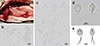 |
Figure 1 Photographs of fresh pseudocysts and myxospores of Henneguya cystigena n. sp. parasitizing the alimentary tract of Acanthopagrus latus. a: yellowish round to oval pseudocysts (arrows) scattered in the stomach wall; b–d: myxospores in frontal and lateral view; e: schematic drawing of a myxospore in frontal (left) and lateral (right) view. |
Description of the new species
Henneguya cystigena n. sp. (Figs. 1–3)
urn:lsid:zoobank.org:act:0FD5F7FB-2F6A-446D-B3AB-CEB40DDC1972
Type host: Yellowfin seabream Acanthopagrus latus (Houttuyn, 1782).
Type locality: East China Sea near the coast of Cangnan county (120°36′39″E, 27°23′36″N), Zhejiang province, China.
Type material: Myxospores preserved in 80% ethanol, National Zoological Museum of China, Institute of Zoology, Chinese Academy of Sciences (IZCAS), collection number NBUECS241102 (syntypes). School of Marine Sciences, Ningbo University, collection number NBUECS241102-1 (syntypes).
Infection site: alimentary tract wall.
Prevalence: 12.5% (1 of 8 fish).
Etymology: the name “cystigena” derives from the generation (from the Latin -gena) of pseudocysts (from the Latin cysti-) by the present species.
Morphology (all measurements in μm): Myxospores 21.7 ± 2.2 (15.9–26.2) in total length. Myxospore body round to ellipsoidal, 9.6 ± 0.5 (8.6–10.6) in length, 7.3 ± 0.4 (6.8–7.9) in width, and 6.0 ± 0.2 (5.5–6.4) in thickness. Two equal-sized pyriform polar capsules located symmetrically in the anterior end of myxospores, measuring 3.5 ± 0.3 (2.9–4.4) × 1.9 ± 0.2 (1.4–2.2) (Figs. 1d–1e). Polar capsules placed at plane parallel to that of sutural line. Two caudal appendages, unequal in length, extending asymmetrically from posterior end, one per valve, one 13.4 ± 1.8 (9.7–16.6) in length, the other 10.2 ± 1.8 (6.4–13.3) in length.
Remarks
We compared phenotypic characters of the present species with those congeners of comparable morphological and morphometric data and parasitizing fish of close relatives (Table 1). All congeners, along with the present species, bear round or ellipsoidal myxospores, with the exception of Henneguya latesi whose myxospore is pyriform. Henneguya lata Chinh et al., 2021 [7] was isolated from the same fish host, but its myxospore width and thickness, and length of polar capsules and caudal appendages are slightly smaller than those of the present species. Henneguya yokoyamai Li et al., 2012 [22], Henneguya ogawai Li et al., 2012 [22], Henneguya cynoscioni Dyková et al., 2011 [12], Henneguya latesi Tripathi, 1952 [35], Henneguya pagri Yokoyama et al., 2005 [38], and Henneguya lateolabracis Yokoyama, et al., 2003 [39] possess longer myxospore length. Compared to the present species, myxospore width and thickness of H. yokoyamai and H. ogawai are smaller, but the measurement of their polar capsules is larger in dimension. Besides, H. cynoscioni, H. pagri, and H. lateolabracis have wider myxospores, but their dimensions of polar capsules are smaller than those of the present species. The caudal appendages of all comparable species here are equal in length, that is distinguishable from those of the present species. Additionally, the lengths of the caudal appendage of H. lata and H. ogawai are smaller and those of other congeners are larger than the present species. Accordingly, the present species differs from all the known congeners and represents a novel species. Therefore, we nominate it as Henneguya cystigena n. sp.
Comparison of morphometric data of Henneguya cystigena n. sp. and its similar congeners.
Molecular comparison
An SSU rDNA sequence comprising 1,932 bp was obtained, with accession number PV221478 available in GenBank. Based on BLASTn search, Henneguya yokoyamai infecting the gall bladder wall of Acanthopagrus schlegelii displayed highest similarity (94.19%, AB693053; Query Cover 100%) to that of the isolate under study. Comparatively, Henneguya lata (MT644624) from the gills of A. latus, and Henneguya ogawai Li et al., 2012 [22] (AB693050) parasitizing the esophageal wall of A. schlegelii shared 93.72% (Query Cover 100%) and 93.15% (Query Cover 100%) molecular similarity, respectively.
Electron microscopy
The observation of SEM showed myxospores exhibiting two smooth shell valves joined along a straight sutural line, with a caudal appendage extending from each valve and tapering posteriorly to the extremity (Figs. 2a and 2b). The results from TEM observation revealed an electron-dense myxospore valve (Figs. 2c–2e). The electron-dense sporoblast was observed inside shell valves, with two capsulogenic cells developing polar tubules. The longitudinal observation of myxospore reveals two polar capsules located anteriorly and one tapered caudal appendage at the posterior portion. Three to four turns of polar tubule cycled inside the polar capsule are observed in an ultra-sliced section.
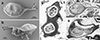 |
Figure 2 Scannning (a–b) and transmission electron microscopy (c–e) images of the myxospores of Henneguya cystigena n. sp. a–b: mxysopres exhibiting two smooth shell valves, with two tapered posterior cadual processes (black arrows). c: two polar capsules (PC) aligned parallel to the plane of sutural line (white arrows); shell valves electron-dense (black arrowheads); d: polar capsules positioned anteriorly, with a single caudal process extending posteriorly from the myxospore body. e: ultrathin transverse sections of a polar capsule revealing four coils of polar tubule (asterisks). |
Histological analysis
Histological sections revealed round to ellipsoidal pseudocysts filled with myxospores developing in the submucous layer of the alimentary tract wall (Fig. 3a). Pseudocysts were entirely encapsulated by the connective tissue, with a peripheral edge confined by a delicate eosinophilic layer (Figs. 3b–3c). An inflammatory reaction was not observed in the surrounding tissue, but a slight distortion of circular muscle layer due to bulging of cysts could be observed. Myxospores inside pseudocysts were observed developing synchronously.
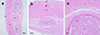 |
Figure 3 Histological sections of pseudocysts (p) formed by Henneguya cystigena n. sp. in the submucosa (s) of the host’s stomach segements (H & E). ad: adventitia; m: circular muscle layer; ms: myxospores. |
Phylogenetic reconstruction
Trees obtained from BI and ML analyses exhibited almost consistent topologies (Fig. 4) and three main clades were artificially defined in the present work. Clade A at the basal position comprises myxosporeans primarily infecting fish belonging to Centrarchiformes and Perciformes. Clade B contains species that mostly parasitize marine fish, including those belonging to the family Sparidae (Eupercaria). Henneguya cystigena n. sp. clustering with H. lata formed a sister group to that composed of H. ogawai and H. yokoyamai in upper Clade B. Lastly, clade C comprises mainly species that infect Siluriformes, but also Characiformes and Esociformes. Species of Henneguya and Myxobolus are intermixed in all three clades.
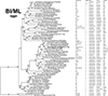 |
Figure 4 Phylogenetic tree reconstructed using Bayesian inference (BI) based on SSU rDNA sequences of Henneguya cystigena n. sp. and related myxosporeans. Bayesian posterior probability and maximum likelihood bootstrap values > 70 are indicated at each node. Dashes represent support values below 70. The species under study is highlighted in bold. For each organism, the following information is provided: GenBank accession number, infection site, habitat type (F: freshwatar; B: brackish water; M: marine; N/A: data not available), host affinity at order and family level, and host sampling location. |
Discussion
The genus Henneguya represents one of the most speciose group within Myxosporea, exhibiting a global distribution and often displaying opportunistic pathogenicity in its fish host. In China, to the best of our knowledge, 35 species of Henneguya have been documented, predominantly from freshwater environments [6, 13, 24, 32, 40, 42]. Among these, Henneguya latesa Wu et al., 1994 isolated from Lates calcarifer (Bloch, 1790) [37] and Henneguya ovata Liu et al., 2018 detected in Trachinotus ovatus (Linnaeus, 1758) [24] are the only two species reported from marine habitats. Consequently, Henneguya cystigena n. sp. described herein represents the third Henneguya species identified along the coast of mainland China.
To ensure accurate species identification, we characterized the present species using a series of microscopic examinations. The observation of a pair of polar capsules lying on the plane parallel to the sutural line, coupled with a single caudal appendage extending from each shell valve, conforms to the taxonomic definition of the genus Henneguya [28]. While Henneguya lata, H. ogawai, and H. yokoyamai share Acanthopagrus fish hosts [7, 22], their myxospore dimensions differ from those of the present species. Furthermore, congeners exhibiting high morphometric resemblance are distinguished from the current isolate by divergence in at least one property, such as structure dimensions, infection site, or fish host specificity (Table 1).
The utilization of molecular data from SSU rDNA sequences significantly enhances accurate myxosporean identification and helps resolve taxonomic ambiguities. BLASTn analysis revealed a maximum sequence identity of 94.19% with all other SSU rDNA sequences available in the NCBI database. Although no universally specified threshold defines interspecific variability for myxosporeans, the observed 6% molecular divergence exceeds the approximately 1% difference commonly employed for delimiting species within Myxobolidae [7, 23, 40, 41]. Based on this substantial molecular distinction, in conjunction with morphological differences, we are confident in designating the present isolate as a novel species, named Henneguya cystigena n. sp.
The genus Henneguya is traditionally distinguished from Myxobolus by the presence of posterior caudal appendages [28]. However, in our phylogenetic reconstruction, Henneguya species appear intermixed with those of Myxobolus across all three main clades. Specifically, H. cystigena n. sp. forms a sister group to a cluster composed of Myxobolus species within clade B. This finding is consistent with previous studies [1, 16, 26] and supports the non-monophyletic origin of the genera Myxobolus and Henneguya, thereby challenging the reliability of caudal appendages as the sole diagnostic character for discriminating between these taxa. Additionally, the clustering patterns observed in our tree corroborate earlier phylogenetic analyses [1, 16, 18, 25, 26, 36, 41], providing further support for the coevolutionary history between these myxosporeans and their fish hosts. Signals related to habitat type, and infection site also exhibit some degree of congruence with grouping of the concerned species, consistent with previous findings [24, 26]. Notably, species within clade A and C primarily parasitize the fish body surface and gills, whereas those in clade B are mostly isolated from internal abdominal organs. This pattern suggests that ancestor of myxosporeans in clade B may have partially undergone an evolutionary adaptation from an original surface-infecting tropism to parasitizing internal organs.
Conclusion
Combining morphological, ultrastructural, and molecular analysis, our results provide robust evidence for the description of a novel Henneguya species parasitizing the alimentary tract wall of wild yellowfin seabream Acanthopagrus latus. Phylogenetic analyses revealed intermixed clustering of myxobolid species, highlighting significant discrepancies between current morphological taxonomy and increasingly refined molecular phylogenies. Furthermore, this study expands our understanding of piscine parasite fauna in marine environments adjacent to mainland China.
Funding
This work was supported by the Ningbo International Science and Technology Cooperation Project (Grant No. 2023H015) and the National Natural Science Foundation of China (Grant No. 32400358).
Conflicts of interest
All authors declare that they have no conflict of interest or competing interests.
Author contribution statement
Bo Zhang: Conceptualization, Methodology, Investigation, Formal analysis, Writing – original draft, Writing – Reviewing & Editing, and Funding acquisition; Fei Yin: Supervision, Resources, Writing – Reviewing & Editing, and Funding acquisition.
References
- Abrunhosa J, Sindeaux-Neto JL, Hamoy I, Matos ER. 2018. A new species of Myxosporea, Henneguya quelen, from silver catfish Rhamdia quelen (Siluriforme: Pimelodidae) in the Amazonian region. Parasitology Research, 117(12), 3809–3820. [Google Scholar]
- Atkinson SD, Bartholomew JL, Lotan T. 2018. Myxozoans: Ancient metazoan parasites find a home in phylum Cnidaria. Zoology, 129, 66–68. [Google Scholar]
- Barta JR, Martin DS, Liberator PA, Dashkevicz M, Anderson JW, Feighner SD, Elbrecht A, PerkinsBarrow A, Jenkins MC, Danforth HD, Ruff MD, ProfousJuchelka H. 1997. Phylogenetic relationships among eight Eimeria species infecting domestic fowl inferred using complete small subunit ribosomal DNA sequences. Journal of Parasitology, 83(2), 262–271. [CrossRef] [Google Scholar]
- Burland TG. 2000. DNASTAR’s Lasergene Sequence Analysis Software, in Bioinformatics Methods and Protocols. Methods in Molecular BiologyTM, Misener SKrawetz SA, Editors. Humana Press, Totowa, NJ, USA, p. 71–91. [Google Scholar]
- Castresana J. 2000. Selection of conserved blocks from multiple alignments for their use in phylogenetic analysis. Molecular Biology and Evolution, 17(4), 540–552. [PubMed] [Google Scholar]
- Chen CL, Ma CL. 1998. Myxozoa: Myxosporea, Science Press, Beijing. [Google Scholar]
- Chinh NN, Ngo HD, Van Tuc V, Itoh N, Yoshinaga T, Shirakashi S, Doanh PN. 2021. A new myxosporean species, Henneguya lata n. sp. (Myxozoa: Myxobolidae), from the gills of yellowfin seabream Acanthopagrus latus (Perciformes: Sparidae) in the Gulf of Tonkin, Vietnam. Parasitology Research, 120(3), 877–885. [CrossRef] [PubMed] [Google Scholar]
- Darriba D, Taboada GL, Doallo R, Posada D. 2012. jModelTest 2: More models, new heuristics and high-performance computing. Nature Methods, 9(8), 772. [CrossRef] [Google Scholar]
- Dereeper A, Audic S, Claverie J-M, Blanc G. 2010. BLAST-EXPLORER helps you building datasets for phylogenetic analysis. BMC Evolutionary Biology, 10, 8. [Google Scholar]
- Dereeper A, Guignon V, Blanc G, Audic S, Buffet S, Chevenet F, Dufayard JF, Guindon S, Lefort V, Lescot M, Claverie JM, Gascuel O. 2008. Phylogeny.fr: robust phylogenetic analysis for the non-specialist, Nucleic Acids Research, 36, W465–W469. [Google Scholar]
- Diep Thi H, Chernomor O, von Haeseler A, Minh BQ, Le Sy V. 2018. UFBoot2: improving the ultrafast bootstrap approximation. Molecular Biology and Evolution, 35(2), 518–522. [CrossRef] [PubMed] [Google Scholar]
- Dyková I, de Buron I, Roumillat WA, Fiala I. 2011. Henneguya cynoscioni sp. n. (Myxosporea: Bivalvulida), an agent of severe cardiac lesions in the spotted seatrout, Cynoscion nebulosus (Teleostei: Sciaenidae). Folia Parasitologica, 58(3), 169–177. [Google Scholar]
- Eiras JC. 2002. Synopsis of the species of the genus Henneguya Thélohan, 1892 (Myxozoa: Myxosporea: Myxobolidae). Systematic Parasitology, 52(1), 43–54. [Google Scholar]
- Eiras JC, Adriano EA. 2012. A checklist of new species of Henneguya Thelohan, 1892 (Myxozoa: Myxosporea, Myxobolidae) described between 2002 and 2012. Systematic Parasitology, 83(2), 95–104. [Google Scholar]
- Fujita T. 1924. Studies on myxosporidian infection of the crucian carp. Japanese Journal of Zoology, 1, 45–47. [Google Scholar]
- Gupta A, Kaur H. 2018. 18S and 28S rDNA identity and phylogeny of two novel myxosporeans infecting gills of cyprinid carps inhabiting a cold water wetland in northern India. Microbial Pathogenesis, 120, 97–108. [Google Scholar]
- Iwatsuki Y. 2013. Review of the Acanthopagrus latus complex (Perciformes: Sparidae) with descriptions of three new species from the Indo-West Pacific Ocean. Journal of Fish Biology, 83(1), 64–95. [Google Scholar]
- Katharios P, Varvarigos P, Keklikoglou K, Ruetten M, Sojan J, Akter M, Cascarano MC, Tsertou MI, Kokkari C. 2020. Native parasite affecting an introduced host in aquaculture: cardiac henneguyosis in the red seabream Pagrus major Temminck & Schlegel (Perciformes: Sparidae) caused by Henneguya aegea n. sp. (Myxosporea: Myxobolidae). Parasites & Vectors, 13(1), 27. [Google Scholar]
- Katoh K, Standley DM. 2013. MAFFT multiple sequence alignment software version 7: Improvements in performance and usability. Molecular Biology and Evolution, 30(4), 772–780. [CrossRef] [Google Scholar]
- Kent ML, Andree KB, Bartholomew JL, El-Matbouli M, Desser SS, Devlin RH, Feist SW, Hedrick RP, Hoffmann RW, Khattra J, Hallett SL, Lester RJ, Longshaw M, Palenzeula O, Siddall ME, Xiao C. 2001. Recent advances in our knowledge of the Myxozoa. Journal of Eukaryotic Microbiology, 48(4), 395–413. [CrossRef] [PubMed] [Google Scholar]
- Klokacheva S. 1914. Ueber die Myxosporidien der Karausche. Zoologischer Anzeiger, 44, 182–186. [Google Scholar]
- Li YC, Sato H, Kamata Y, Ohnishi T, Sugita-Konishi Y. 2012. Three novel myxobolid species of genera Henneguya and Myxobolus (Myxosporea: Bivalvulida) from marine fish in Japan. Parasitology Research, 111(2), 819–826. [Google Scholar]
- Lisnerová M, Blabolil P, Holzer A, Jurajda P, Fiala I. 2020. Myxozoan hidden diversity: the case of Myxobolus pseudodispar Gorbunova, 1936. Folia Parasitologica, 67, 019. [Google Scholar]
- Liu XH, Xu LW, Luo D, Zhao YL, Liu GF, Zhang QQ, Zhang JY. 2018. Henneguya ovata n.sp. (Myxosporea: Bivalvulida), causing severe enteric henneguyosis of net-cage-cultured ovate pompano, Trachinotus ovatus in China. Aquaculture, 483, 8–15. [Google Scholar]
- Liu XH, Zhang JY, Batueva MD, Voronin VN. 2016. Supplemental description and molecular characterization of Myxobolus miyarii Kudo, 1919 (Myxosporea: Myxobolidae) infecting intestine of Amur catfish (Silurus asotus). Parasitology Research, 115(4), 1547–1556. [Google Scholar]
- Liu Y, Lovy A, Gu Z, Fiala I. 2019. Phylogeny of Myxobolidae (Myxozoa) and the evolution of myxospore appendages in the Myxobolus clade. International Journal for Parasitology, 49(7), 523–530. [CrossRef] [PubMed] [Google Scholar]
- Lom J, Arthur JR. 1989. A guideline for the preparation of species descriptions in Myxosporea. Journal of Fish Diseases, 12(2), 151–156. [CrossRef] [Google Scholar]
- Lom J, Dyková I. 2006. Myxozoan genera: Definition and notes on taxonomy, life-cycle terminology and pathogenic species. Folia Parasitologica, 53(1), 1–36. [CrossRef] [PubMed] [Google Scholar]
- Nguyen LT, Schmidt HA, von Haeseler A, Minh BQ. 2015. IQ-TREE: a fast and effective stochastic algorithm for estimating maximum-likelihood phylogenies. Molecular Biology and Evolution, 32(1), 268–274. [CrossRef] [PubMed] [Google Scholar]
- Okamura B, Hartigan A, Naldoni J. 2018. Extensive uncharted biodiversity: the parasite dimension. Integrative and Comparative Biology, 58(6), 1132–1145. [PubMed] [Google Scholar]
- Pote LMW, Khoo L, Griffin M. 2012. Henneguya ictaluri, in Fish Parasites: Pathobiology and Protection, Woo PTK, Buchmann K, Editors. CABI, Wallingford, Oxfordshire, UK, p. 177–192. [Google Scholar]
- Rangel LF, Santos MJ, Rocha S. 2023. Synopsis of the species of Henneguya Thelohan, 1892 (Cnidaria: Myxosporea: Myxobolidae) described since 2012. Systematic Parasitology, 100(3), 291–305. [Google Scholar]
- Ronquist F, Teslenko M, van der Mark P, Ayres DL, Darling A, Höhna S, Larget B, Liu L, Suchard MA, Huelsenbeck JP. 2012. MrBayes 3.2: Efficient Bayesian phylogenetic inference and model choice across a large model space. Systematic Biology, 61(3), 539–542. [CrossRef] [PubMed] [Google Scholar]
- Stilwell JM, Camus AC, Leary JH, Khoo LH, Griffin MJ. 2019. Pathologic changes associated with respiratory compromise and morbidity due to massive interlamellar Henneguya exilis infection in Channel × Blue Hybrid Catfish. Journal of Parasitology, 105(5), 686–692. [Google Scholar]
- Tripathi YR. 1952. Studies on parasites of Indian fishes, I, Protozoa Myxosporidia together with a check list of parasitic protozoa described from Indian fishes. Records of the Indian Museum, 50, 63–88. [Google Scholar]
- Velasco M, Videira M, Silva do Nascimento, Matos, P., Goncalves, E.C., Matos, E.LDC, Matos P, Goncalves EC, MatosE. 2016. Henneguya paraensis n. sp. (Myxozoa; Myxosporea), a new gill parasite of the Amazonian fish Cichla temensis (Teleostei: Cichlidae): morphological and molecular aspects. Parasitology Research, 115(5), 1779–1787. [Google Scholar]
- Wu Z, Wu J. 1994. A study on Myxosporidia of fishes from South China Sea. Tropic Oceanology, 13(3), 67–71. [Google Scholar]
- Yokoyama H, Itoh N, Tanaka S. 2005. Henneguya pagri n. sp. (Myxozoa: Myxosporea) causing cardiac henneguyosis in red sea bream, Pagrus major (Temminck & Schlegel). Journal of Fish Diseases, 28(8), 479–487. [Google Scholar]
- Yokoyama H, Kawakami H, Yasuda H, Tanaka S. 2003. Henneguya lateolabracis sp. n. (Myxozoa : Myxosporea), the causative agent of cardiac henneguyosis in Chinese sea bass Lateolabrax sp. Fisheries Science, 69(6), 1116–1120. [Google Scholar]
- Yuan S, Xu LW, Weng MQ, Zhang JY, Sato H. 2021. Outbreak of gill henneguyosis in pond-cultured Oxyeleotris spp. (Eleotridae, Perciformes) caused by Henneguya shaharini Shariff, 1982 (Myxosporea: Bivalvulida) in South China, Aquaculture, 538, 736587. [Google Scholar]
- Zatti SA, Atkinson SD, Maia AAM, Bartholomew JL, Adriano EA. 2018. Novel Henneguya spp. (Cnidaria: Myxozoa) from cichlid fish in the Amazon basin cluster by geographic origin. Parasitology Research, 117(3), 849–859. [Google Scholar]
- Zhang B, Tu X, Gu ZM. 2023. Henneguyosis: A novel threat to the exotic channel catfish Ictalurus punctatus cultivated in China. Aquaculture, 576, 739831. [Google Scholar]
Cite this article as: Zhang B & Yin F. 2025. Morphological and molecular characterization of Henneguya cystigena n. sp. (Cnidaria, Myxosporea) parasitizing the alimentary tract of yellowfin seabream, Acanthopagrus latus, in the East China Sea. Parasite 32, 51. https://doi.org/10.1051/parasite/2025048.
All Tables
Comparison of morphometric data of Henneguya cystigena n. sp. and its similar congeners.
All Figures
|
Figure 1 Photographs of fresh pseudocysts and myxospores of Henneguya cystigena n. sp. parasitizing the alimentary tract of Acanthopagrus latus. a: yellowish round to oval pseudocysts (arrows) scattered in the stomach wall; b–d: myxospores in frontal and lateral view; e: schematic drawing of a myxospore in frontal (left) and lateral (right) view. |
| In the text | |
|
Figure 2 Scannning (a–b) and transmission electron microscopy (c–e) images of the myxospores of Henneguya cystigena n. sp. a–b: mxysopres exhibiting two smooth shell valves, with two tapered posterior cadual processes (black arrows). c: two polar capsules (PC) aligned parallel to the plane of sutural line (white arrows); shell valves electron-dense (black arrowheads); d: polar capsules positioned anteriorly, with a single caudal process extending posteriorly from the myxospore body. e: ultrathin transverse sections of a polar capsule revealing four coils of polar tubule (asterisks). |
| In the text | |
|
Figure 3 Histological sections of pseudocysts (p) formed by Henneguya cystigena n. sp. in the submucosa (s) of the host’s stomach segements (H & E). ad: adventitia; m: circular muscle layer; ms: myxospores. |
| In the text | |
|
Figure 4 Phylogenetic tree reconstructed using Bayesian inference (BI) based on SSU rDNA sequences of Henneguya cystigena n. sp. and related myxosporeans. Bayesian posterior probability and maximum likelihood bootstrap values > 70 are indicated at each node. Dashes represent support values below 70. The species under study is highlighted in bold. For each organism, the following information is provided: GenBank accession number, infection site, habitat type (F: freshwatar; B: brackish water; M: marine; N/A: data not available), host affinity at order and family level, and host sampling location. |
| In the text | |
Current usage metrics show cumulative count of Article Views (full-text article views including HTML views, PDF and ePub downloads, according to the available data) and Abstracts Views on Vision4Press platform.
Data correspond to usage on the plateform after 2015. The current usage metrics is available 48-96 hours after online publication and is updated daily on week days.
Initial download of the metrics may take a while.