Figure 2
Download original image
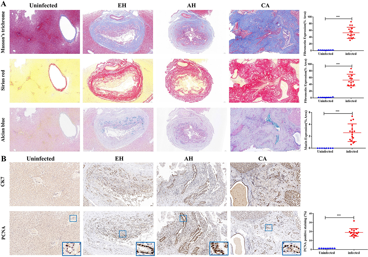
The histopathological examination of hepatobiliary tissues in cats. A: Histopathological analysis using Masson’s trichrome, Sirius red, and Alcian blue staining techniques to analyze the intrahepatic bile duct tissues of cats, including the analysis of the area proportion of positive staining. B: Immunohistochemical staining of intrahepatic bile duct tissues of cats using CK7 and PCNA, analyzing the IOD/Area value for PCNA positive expression. The samples were examined at 200× magnifications. Statistical significance: *p < 0.05; **p < 0.01; ***p < 0.005; ****p < 0.001.
Current usage metrics show cumulative count of Article Views (full-text article views including HTML views, PDF and ePub downloads, according to the available data) and Abstracts Views on Vision4Press platform.
Data correspond to usage on the plateform after 2015. The current usage metrics is available 48-96 hours after online publication and is updated daily on week days.
Initial download of the metrics may take a while.