| Issue |
Parasite
Volume 30, 2023
|
|
|---|---|---|
| Article Number | 51 | |
| Number of page(s) | 8 | |
| DOI | https://doi.org/10.1051/parasite/2023051 | |
| Published online | 28 November 2023 | |
Research Article
First report of prevalence and assemblage analysis of Giardia duodenalis in pigs from Guangxi Zhuang Autonomous Region, southern China
Premier rapport sur la prévalence et l’analyse des assemblages de Giardia duodenalis chez les porcs de la région autonome Zhuang du Guangxi, dans le sud de la Chine
1
Guangxi Vocational University of Agriculture, Nanning 530007, China
2
Key Laboratory of Fujian-Taiwan Animal Pathogen Biology, College of Animal Sciences (College of Bee Science), Fujian Agriculture and Forestry University, Fuzhou 350002, China
* Corresponding authors: This email address is being protected from spambots. You need JavaScript enabled to view it.
; This email address is being protected from spambots. You need JavaScript enabled to view it.
Received:
21
August
2023
Accepted:
3
November
2023
Abstract
Giardia duodenalis is a common intestinal protozoan that can cause diarrhea and intestinal disease in animals and in humans. However, the prevalence and assemblages of G. duodenalis in pigs from Guangxi Zhuang Autonomous Region have not been reported. In this study, a total of 724 fecal samples (201 from nursery pigs, 183 from piglets, 175 from breeding pigs, and 165 from fattening pigs) were obtained in four areas of the region (Nanning, Yulin, Hezhou, and Guigang). The gene of the small subunit ribosomal RNA (SSU rRNA) of G. duodenalis was amplified by nested PCR. The results show that the prevalence of G. duodenalis in pigs was 3.59% (26/724), of which 14 samples belonged to assemblage A (53.85%) and 12 samples belonged to assemblage E (46.15%). The infection rates of G. duodenalis in Hezhou, Yulin, Nanning, and Guigang were 0%, 0.7%, 10.8% and 1.1%, respectively (χ2 = 45.616, p < 0.01); whereas 5.1% of breeding pigs, 6.0% of piglets, 2.4% of fattening pigs, and 1.0% of nursery pigs were infected with G. duodenalis (χ2 = 8.874, p < 0.05). The SSU rRNA-positive samples were amplified by PCR based on the β-giardin (bg), glutamate dehydrogenase (gdh), and triphosphate isomerase (tpi) genes. Ten, eight and seven positive samples were detected, respectively. Based on phylogenetic analysis of the three genetic loci sequences, a multilocus genotyping A1 was found. The findings of this study provide basic data for the development of prevention and control of G. duodenalis infections in pigs and humans in the Guangxi Zhuang Autonomous Region.
Résumé
Giardia duodenalis est un protozoaire intestinal commun qui peut provoquer des diarrhées et des maladies intestinales chez les animaux et les humains. Cependant, la prévalence et les assemblages de G. duodenalis chez les porcs de la région autonome Zhuang du Guangxi n’ont pas été rapportés. Dans cette étude, un total de 724 échantillons fécaux (201 provenant de jeunes porcelets, 183 de porcelets, 175 de porcs reproducteurs et 165 de porcs à l’engrais) ont été obtenus dans quatre zones de la région (Nanning, Yulin, Hezhou et Guigang). Le gène de la petite sous-unité de l’ARN ribosomal (ARNr SSU) de G. duodenalis a été amplifié par PCR nichée. Les résultats ont montré que la prévalence de G. duodenalis chez les porcs était de 3,59 % (26/724), dont 14 échantillons appartenaient à l’assemblages A (53,85 %) et 12 échantillons à l’assemblage E (46,15 %). Les taux d’infection par G. duodenalis à Hezhou, Yulin, Nanning et Guigang étaient respectivement de 0, 0,7 %, 10,8 % et 1,1 % (χ2 = 45,616, p < 0,01), alors que 5,1 % des porcs reproducteurs, 6,0 % des porcelets, 2,4 % de porcs à l’engrais et 1,0 % des jeunes porcelets étaient infectés par G. duodenalis (χ2 = 8,874, p < 0,05). Les échantillons positifs pour l’ARNr SSU ont été amplifiés par PCR basée sur les gènes de la β-giardine (bg), de la glutamate déshydrogénase (gdh) et de la triphosphate isomérase (tpi), et dix, huit et sept échantillons positifs ont été détectés, respectivement. Sur la base de l’analyse phylogénétique des trois séquences de loci génétiques, un génotypage multilocus A1 a été trouvé. Les résultats de cette étude fournissent des données de base pour le développement de la prévention et du contrôle des infections à G. duodenalis chez les porcs et les humains dans la région autonome Zhuang du Guangxi.
Key words: Giardia duodenalis / Pigs / Prevalence / Multilocus genotyping / Guangxi Zhuang Autonomous Region
Edited by Jean-Lou Justine
© Y.-F. Song et al., published by EDP Sciences, 2023
 This is an Open Access article distributed under the terms of the Creative Commons Attribution License (https://creativecommons.org/licenses/by/4.0), which permits unrestricted use, distribution, and reproduction in any medium, provided the original work is properly cited.
Introduction
Giardia spp. are common intestinal parasites, affecting both humans and a wide variety of other animals [13]. The parasite was first discovered over 300 years ago by Antonie van Leeuwenhoek, and since then, six distinct Giardia species have been described [9]. The six described Giardia species (Giardia agilis, Giardia psittaci, Giardia ardeae, Giardia muris, Giardia microti, and Giardia duodenalis) infect a wide range of animals including birds, amphibians, rodents, and mammals [20]. Giardia duodenalis is an important zoonotic parasite that can cause giardiasis [32, 35]. The life cycle of G. duodenalis is simple. It consists of two key stages: rapidly multiplying trophozoites which attach to intestinal epithelial cells, and cysts with high resistance to environmental degradation, that are released in the feces and spread through the fecal-oral route [11, 19, 23]. Infection with G. duodenalis shows a wide range of clinical symptoms, such as acute or chronic diarrhea, nausea, abdominal pain, vomiting, and weight loss [10, 39]. Furthermore, giardiasis affects growth, development, and cognitive functions in infected children [13, 34].
The comprehension of G. duodenalis through molecular biological analysis has greatly contributed to the understanding of its taxonomy, population genetics, and epidemiology. The small subunit ribosomal RNA (SSU rRNA), β-giardin (bg), glutamate dehydrogenase (gdh), and triose phosphate isomerase (tpi) genes are four commonly used gene loci in the genotyping of G. duodenalis, but a single gene may not correctly identify G. duodenalis or completely describe its genetic characterization [25, 43]. Multilocus genotyping (MLG) based on more than three genes is considered to be more reliable than single-locus genotyping in assemblage and sub-assemblage typing of isolates. As a result, it has been widely used to investigate the genotypic diversity of G. duodenalis, and is considered to be useful for detecting and identifying mixed infections by different assemblages of the parasite [13, 37]. To date, G. duodenalis can be divided into eight assemblages (A–H) based on genetic analysis, with each assemblage exhibiting a distinct host range [5, 20, 27]. Among the assemblages, assemblages A and B can be found in a wide array of mammals, including humans and pigs [13, 31]. Other assemblages (C–H) exhibit host specificity and narrow host ranges: assemblages C and D are specific to dogs and other canids; assemblage E is found in domestic animals, such as pigs, cattle, and horses; assemblage F has been identified in cats; assemblages G and H are found in rodents and marine animals such as seals, respectively [8, 13, 26]. However, assemblages C–F have also been identified in humans [22, 29, 35, 44]. These findings suggest that close contact between humans and animals may lead to human infection with G. duodenalis.
Although some studies have reported prevalence and distribution of G. duodenalis in pigs [1, 15, 17, 18, 41], there are no reports on the infection of G. duodenalis in pigs in Guangxi Zhuang Autonomous Region, China. Pigs infected with G. duodenalis may cause malabsorption and weight loss, resulting in a decline in pig production [48]. Currently, assemblages A–F have been found in pigs, with assemblage E the preponderant genotype [4]. Therefore, this study aimed to investigate the prevalence of G. duodenalis infection and identify the genotypes present in pigs from Guangxi Zhuang Autonomous Region, China. The study provides essential data concerning G. duodenalis infections in pigs in southern China, which can contribute to the development of targeted public health and effective strategies for prevention and control of giardiasis in this area.
Materials and methods
Samples collection
From March 2021 to May 2022, a total of 724 fecal samples (201 from nursery pigs, 183 from piglets, 175 from breeding pigs, and 165 from fattening pigs) were collected from four cities (Nanning, Yulin, Hezhou, and Guigang) in Guangxi Zhuang Autonomous Region, China (Fig. 1). Fecal samples were collected directly from the rectum by using sterile gloves. Subsequently, fecal samples were placed in sterile plastic bags marked with the date, type and farm. All samples were promptly transported to the laboratory in cool boxes with ice packs and stored at −80 °C until DNA extraction.
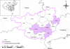 |
Figure 1 Map of the sample collection area. The green areas are the four areas where samples were collected in Guangxi Zhuang Autonomous Region, China. |
Genomic DNA extraction
Approximately 200 mg of each fecal sample was aseptically transferred to individual 1.5 mL centrifuge tubes. Genomic DNA extraction was performed using an E.Z.N.A.® Stool DNA Kit (D4015-02, OMEGA Bio-Tek Inc., Norcross, GA, USA), according to the manufacturer’s instructions. The extracted DNA was subsequently stored at −20 °C to maintain its integrity for subsequent analyses.
PCR amplification
All samples were amplified by using nested PCR targeting the SSU rRNA gene, as previously described [2]. The PCR reaction mixture comprising 25 μL was prepared and amplified according to the procedure described by Jing et al. [15]. All SSU rRNA-positive samples were subjected to amplification using nested PCR targeting the bg, gdh, and tpi genes, as previously described [7, 16, 36]. A positive control (DNA from G. duodenalis stored in the laboratory at −80 °C) and a negative control (distilled water) were included in each PCR assay. Each sample was processed three times at the SSU rRNA gene and each SSU rRNA-positive sample was processed three times at the bg, gdh, and tpi genes. The PCR products were identified by 1.5% agarose gel electrophoresis (Gene Biotechnology International Trade Co., Ltd., Shanghai, China).
Sequencing and phylogenetic analysis
PCR products of positive samples were processed for two-directional sequencing by Sangon Biotech (Xiamen, China). The obtained DNA sequences were aligned with homologous sequences available in the GenBank database of the National Center for Biotechnology Information (https://www.ncbi.nlm.nih.gov/). The MLG method was only used to analyze the genetic diversity of positive samples which were successfully sequenced at all three gene loci (bg, gdh, and tpi). Neighbor-joining trees [33] were constructed using Mega 11 software, employing the kimura-2 parameter model. The reliability of these trees was assessed using the bootstrap method with 1,000 pseudoreplicates.
Statistical analysis
The Chi-square test (SPSS 25.0 Inc., Chicago, IL, USA) was used to assess differences in prevalence between regions, feeding stages and farming practices. Differences of p < 0.05 were considered statistically significant, and differences of p < 0.01 were considered extremely significant.
Nucleotide sequence accession numbers
All nucleotide sequences have been submitted to the GenBank database in NCBI and allocated accession numbers as follows: OQ943958–OQ943959 for the SSU rRNA gene, OQ934094–OQ934095 for the bg gene, OQ934096–OQ934101 for the gdh gene, and OQ934102–OQ934104 for the tpi gene.
Results
Out of 724 fecal samples collected from 4 cities, 26 samples (26/724) tested positive for G. duodenalis based on the SSU rRNA gene. The highest infection rate of G. duodenalis was in Nanning (10.8%, 23/213), followed by Guigang (1.1%, 2/177), Yulin (0.7%, 1/146), and Hezhou (0%, 0/188). Statistical analysis demonstrated significant variations in the positive rate of G. duodenalis infection with pigs among different regions (χ2 = 45.616, p < 0.01). Among the various feeding stages, piglets had the highest infection rate (6%, 11/183), followed by breeding pigs (5.1%, 9/175), fattening pigs (2.4%, 4/165), and nursery pigs (1%, 2/201) (χ2 = 8.874, p < 0.05). The prevalence of G. duodenalis infection in intensive and free-range farms were 3.7% (15/410) and 3.5% (11/314), respectively. Statistical analysis showed no significant difference between the two groups (p > 0.05) (Table 1).
Investigation of G. duodenalis infection in pigs in Guangxi Zhuang Autonomous Region.
Sequence analysis of the SSU rRNA gene showed two assemblages (A and E) of G. duodenalis in pigs. Of the 26 SSU rRNA-positive samples, 14 isolates (53.85%, 14/26) belong to zoonotic assemblage A, while the other 12 isolates (46.15%, 12/26) were identified as assemblage E (Table 1). Ten, eight and seven sequences were obtained by amplifying 26 SSU rRNA-positive samples at the bg, gdh, and tpi gene loci, respectively (Table 1). At the bg locus, six and four isolates belong to assemblages A and E, respectively; at the gdh locus, six and two isolates belonged to assemblages A and E, respectively; and at the tpi locus, five and two isolates belonged to assemblages A and E, respectively (Table 1). Notably, only one fecal sample of assemblage A was successfully sequenced at all three gene loci and one MLG A1 was formed (Table 2).
Genotyping of G. duodenalis based on the SSU rRNA, bg, gdh, and tpi genes.
To explore the genetic relationships of the G. duodenalis isolates from pigs, four phylogenetic trees were constructed using the SSU rRNA, bg, gdh, and tpi sequences of the parasite. The results show that the representative isolates at the four gene loci were distributed into assemblages A and E, respectively (Fig. 2).
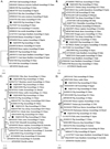 |
Figure 2 The phylogenetic relationships of G. duodenalis isolates were obtained by neighbor-joining analysis. Bootstrap values > 50% from 1,000 replicates are shown as nodes. The black circles represent the sequences obtained in this study. A. Phylogenetic relationships based on SSU rRNA nucleotide sequences; B. Phylogenetic relationships based on bg nucleotide sequences; C. Phylogenetic relationships based on gdh nucleotide sequences; D. Phylogenetic relationships based on tpi nucleotide sequences. |
Discussion
Giardia duodenalis is an intestinal parasitic protozoan that has attracted much attention in recent years. Infection with G. duodenalis is widespread in pigs, affecting individuals of all age groups [24]. In this study, four areas of the Guangxi Zhuang Autonomous Region were investigated, and it was found that the positive rate of G. duodenalis was 3.59% in 724 samples. There have been no reports of pigs infected with G. duodenalis in Vietnam, a neighboring region of the region. However, a report by Verle et al. showed that the prevalence of G. duodenalis infection in humans from Vietnam was 3.2% [38]. Therefore, the results of this study can provide a reference for Vietnam, and further research is needed to determine whether the cause of human infection with G. duodenalis is related to pigs [38]. The infection rates of G. duodenalis in pigs vary around the world, such as 1% (6/633) in Canada, 3.4% (3/90) in Brazil, 14% (120/856) in Denmark, 14.8% (110/745) in Korea, 25.4% (53/209) in Nigeria, and 31.1% (90/289) in Western Australia [1, 3, 6, 12, 17, 28]. In China, the infection rate of G. duodenalis in this investigation was similar to the prevalence in Sichuan Province (3.1%, 11/357) [21]; it was higher than that detected in Hubei Province (0.97%, 8/826), Xinjiang (2.6%, 21/801) and Henan Province (1.7%, 15/897) [15, 18, 40], but lower than that reported in Guangdong Province (18.04%, 94/521), Fujian Province (26.9%, 195/725), Shaanxi Province (8.0%, 45/560), and Zhejiang Province (10.5%, 13/124) [41, 46–48]. These differences can be attributed to several factors, including sample conditions, feeding patterns, age groups, testing methods and seasons changes [4]. We speculate that the strict prevention and control measures implemented after the African swine fever outbreak in China may have resulted in lower infection rates in this study than the rates reported in other regions of China. Moreover, the increased frequency and intensity of disinfection have played a crucial role in reducing the transmission of pathogens.
The positive rate in different regions and for different feeding stages was significantly different (χ2 = 45.616, p < 0.01; χ2 = 8.874, p < 0.05). The highest prevalence (10.8%, 23/213) was observed in Nanning, and the lowest prevalence (0%, 0/188) in Hezhou. Among the different feeding stages, the infection rates of breeding pigs (5.1%, 9/175) and piglets (6.0%, 11/183) were higher than those of fattening pigs (2.4%, 4/165) and nursery pigs (1.0%, 2/201). Similarly, a study in Henan Province found that piglets had higher infection rates (5.8%, 14/243) compared to fattening pigs (0.2%, 1/439) [40]. In contrast, a study in Xinjiang found that the infection rate was highest in fattening pigs (5.4%, 7/129) and lowest in pre-weaned piglets (1.2%, 2/169) [15]. Previous studies have shown that different prevalence in pigs of different age groups may be caused by gut microbiota, nutritional status, immunity and geographical isolation [28]. The higher rate of infection in piglets may be attributed to reduced immunity. During this stage, piglets have not yet fully developed their own immune system [6, 24]. Additionally, factors such as environmental conditions, inadequate nutrition, and stress can further compromise their immune defenses. These situations increase susceptibility to G. duodenalis and other pathogens.
No significant differences were found in the positive rates for different feeding methods in our study (p > 0.05). The positive rate of intensive feeding farms was 3.7% (15/410), while the positive rate of free-range farms was 3.5% (11/314). The low positive rates of both intensive feeding and free-range farms indicates that people have begun to pay attention to the prevention and control of giardiasis. However, the possibility that insufficient samples on free-range farms led to lower infection rates cannot be ruled out.
Giardia duodenalis zoonotic assemblage A (n = 14, 53.85%) and assemblage E (n = 12, 46.15%) were identified among the 26 samples. Assemblage A was the predominant genotype in this study. However, in Australia, Armson et al. identified 56 G. duodenalis positive samples belonging to assemblage E (37 samples, 64.9%) and assemblage A (19 samples, 33.3%) [3]. A study in Denmark found that 13 G. duodenalis positive samples belonged to assemblage E (11 samples, 84.6%) and assemblage A (2 samples, 15.4%) [28]. The finding that pig-derived assemblage A isolates have 100% homology with human-derived isolates at the SSU rRNA locus means the possibility of zoonotic transmission in this areas. Notably, we also identified assemblage E, which has been reported to infect humans [14].
To further expand knowledge of the genetic diversity of G. duodenalis in pigs, the sequence characters of the bg, gdh, and tpi genes were analyzed for the 26 SSU rRNA positive samples and the MLGs were characterized in pigs based on data from these three loci. Amplification success rates at the bg, gdh, and tpi loci varied from 26.92% to 38.46%. The genetic loci of Giardia have different substitution rates, leading to different resolution for parasite typing at varied loci [13]. Nested PCR protocols based on multiple-copy genes (e.g. SSU rRNA) have higher diagnostic sensitivities than those based on single-copy genes (bg, gdh, and tpi) [45]. Therefore, the SSU rRNA-positive samples not amplified at the other three loci may be due to the limited sensitivity of PCR in detecting the single-copy genes. The present study confirmed that more positive samples of G. duodenalis can be amplified based on the SSU rRNA gene. The same problem has been reported in similar studies in pigs in Xinjiang [15]. The results showed that one fecal sample of assemblage A was successfully sequenced at all three loci, with one MLG, A1. In Ogun state of Nigeria, 12 SSU rRNA-positive samples were simultaneously amplified at three loci, with four MLGs [1]. In Shaanxi Province, eight SSU rRNA-positive samples were simultaneously amplified at three loci, with four MLGs [41]. In Fujian Province, six SSU rRNA-positive samples were simultaneously amplified at three loci, with one MLG [46]. In Spain, 76 SSU rRNA-positive samples from humans were simultaneously amplified at three loci, with 23 MLGs [42]. In Southwestern Iran, 82 SSU rRNA-positive samples from humans were simultaneously amplified at three loci, with two MLGs [30]. These results show that the polymorphism of G. duodenalis is different in different regions.
In short, pigs infected with G. duodenalis in Guangxi Zhuang Autonomous Region may be a potential source of infectious cysts that affect humans. Workers on pig farms and in slaughterhouses are at risk of infection with G. duodenalis. Understanding the transmission dynamics between animals and humans is crucial for effective disease control and prevention strategies. Therefore, public health problems caused by G. duodenalis should be investigated.
In conclusion, this study provides the first report of giardiasis in pigs from Guangxi Zhuang Autonomous Region, China. Although a low infection rate of G. duodenalis (3.59%, 26/724) was found in this study, the identification of assemblages A and E, and the predominance of assemblage A, suggest that pigs may play an important role in the transmission of G. duodenalis. The multilocus genotyping results of bg, tpi, and gdh loci showed only one MLG A1. The identification and understanding of the distribution of G. duodenalis in animals, such as pigs, are crucial for the development and implementation of effective prevention and control measures. These findings highlight the importance of continued surveillance and targeted interventions to mitigate the risk of G. duodenalis transmission between animals and humans. The data presented in this study serve as a foundation for future research on G. duodenalis.
Conflict of interest
All authors declare that they have no conflict of interest.
Acknowledgments
This work was supported by the Guangxi Natural Science Foundation (Grant No. 2019GXNSFBA185009).
References
- Akinkuotu OA, Takeet MI, Otesile EB, Olufemi F, Greenwood SJ, McClure JT. 2019. Prevalence and multilocus genotypes of Giardia duodenalis infecting pigs in Ogun state, Nigeria. Infection, Genetics and Evolution: Journal of Molecular Epidemiology and Evolutionary Genetics in Infectious Diseases, 70, 53–60. [CrossRef] [Google Scholar]
- Appelbee AJ, Frederick LM, Heitman TL, Olson ME. 2003. Prevalence and genotyping of Giardia duodenalis from beef calves in Alberta, Canada. Veterinary Parasitology, 112(4), 289–294. [CrossRef] [PubMed] [Google Scholar]
- Armson A, Yang R, Thompson J, Johnson J, Reid S, Ryan UM. 2009. Giardia genotypes in pigs in Western Australia: prevalence and association with diarrhea. Experimental Parasitology, 121(4), 381–383. [CrossRef] [PubMed] [Google Scholar]
- Asghari A, Ebrahimi M, Shamsi L, Sadrebazzaz A, Shams M. 2023. Global molecular prevalence of Giardia duodenalis in pigs (Sus domesticus): A systematic review and meta-analysis. Heliyon, 9(2), e13243. [CrossRef] [PubMed] [Google Scholar]
- Bartelt LA, Sartor RB. 2015. Advances in understanding Giardia: determinants and mechanisms of chronic sequelae. F1000 Prime Reports, 7, 62. [Google Scholar]
- Budu-Amoako E, Greenwood SJ, Dixon BR, Barkema HW, Hurnik D, Estey C, McClure JT. 2012. Occurrence of Giardia and Cryptosporidium in pigs on Prince Edward Island, Canada. Veterinary Parasitology, 184(1), 18–24. [CrossRef] [PubMed] [Google Scholar]
- Cacciò SM, Beck R, Lalle M, Marinculic A, Pozio E. 2008. Multilocus genotyping of Giardia duodenalis reveals striking differences between assemblages A and B. International Journal for Parasitology, 38(13), 1523–1531. [CrossRef] [PubMed] [Google Scholar]
- de Aquino MCC, Inácio SV, Rodrigues FS, de Barros LD, Garcia JL, Headley SA, Gomes JF, Bresciani KDS. 2020. Cryptosporidiosis and Giardiasis in buffaloes (Bubalus bubalis). Frontiers in Veterinary Science, 7. [Google Scholar]
- Dobell C. 1920. The discovery of the intestinal protozoa of Man. Proceedings of the Royal Society of Medicine, 13 (Sect Hist Med), 1–15. [CrossRef] [PubMed] [Google Scholar]
- Eckmann L. 2003. Mucosal defences against Giardia. Parasite Immunology, 25(5), 259–270. [CrossRef] [PubMed] [Google Scholar]
- Einarsson E, Ma’ayeh S, Svärd SG. 2016. An up-date on Giardia and giardiasis. Current Opinion in Microbiology, 34, 47–52. [CrossRef] [PubMed] [Google Scholar]
- Fava NM, Soares RM, Scalia LA, Kalapothakis E, Pena IF, Vieira CU, Faria ES, Cunha MJ, Couto TR, Cury MC. 2013. Performance of glutamate dehydrogenase and triose phosphate isomerase genes in the analysis of genotypic variability of isolates of Giardia duodenalis from livestocks. Biomed Research International, 2013, 875048. [PubMed] [Google Scholar]
- Feng Y, Xiao L. 2011. Zoonotic potential and molecular epidemiology of Giardia species and giardiasis. Clinical Microbiology Reviews, 24(1), 110–140. [CrossRef] [PubMed] [Google Scholar]
- Foronda P, Bargues MD, Abreu-Acosta N, Periago MV, Valero MA, Valladares B, Mas-Coma S. 2008. Identification of genotypes of Giardia intestinalis of human isolates in Egypt. Parasitology Research, 103(5), 1177–1181. [CrossRef] [PubMed] [Google Scholar]
- Jing B, Zhang Y, Xu C, Li D, Xing J, Tao D, Zhang L, Qi M, Wang H. 2019. Detection and genetic characterization of Giardia duodenalis in pigs from large-scale farms in Xinjiang, China. Parasite, 26, 53. [CrossRef] [EDP Sciences] [PubMed] [Google Scholar]
- Lalle M, Pozio E, Capelli G, Bruschi F, Crotti D, Cacciò SM. 2005. Genetic heterogeneity at the beta-giardin locus among human and animal isolates of Giardia duodenalis and identification of potentially zoonotic subgenotypes. International Journal for Parasitology, 35(2), 207–213. [CrossRef] [PubMed] [Google Scholar]
- Lee H, Jung B, Lim JS, Seo MG, Lee SH, Choi KH, Hwang MH, Kim TH, Kwon OD, Kwak D. 2020. Multilocus genotyping of Giardia duodenalis from pigs in Korea. Parasitology International, 78, 102154. [CrossRef] [PubMed] [Google Scholar]
- Li D, Deng H, Zheng Y, Zhang H, Wang S, He L, Zhao J. 2022. First characterization and zoonotic potential of Cryptosporidium spp. and Giardia duodenalis in pigs in Hubei Province of China. Frontiers in Cellular and Infection. Microbiology, 12, 949773. [Google Scholar]
- Li J, Qin H, Li X, Zhang L. 2023. Role of rodents in the zoonotic transmission of giardiasis. One Health, 16, 100500. [CrossRef] [PubMed] [Google Scholar]
- Li J, Wang H, Wang R, Zhang L. 2017. Giardia duodenalis Infections in humans and other animals in China. Frontiers in Microbiology, 8, 2004. [CrossRef] [PubMed] [Google Scholar]
- Li W, Deng L, Wu K, Huang X, Song Y, Su H, Hu Y, Fu H, Zhong Z, Peng G. 2017. Presence of zoonotic Cryptosporidium scrofarum, Giardia duodenalis assemblage A and Enterocytozoon bieneusi genotypes in captive Eurasian wild boars (Sus scrofa) in China: potential for zoonotic transmission. Parasites & Vectors, 10(1), 10. [CrossRef] [PubMed] [Google Scholar]
- Liu H, Shen Y, Yin J, Yuan Z, Jiang Y, Xu Y, Pan W, Hu Y, Cao J. 2014. Prevalence and genetic characterization of Cryptosporidium, Enterocytozoon, Giardia and Cyclospora in diarrheal outpatients in China. BMC Infectious Diseases, 14, 25. [CrossRef] [PubMed] [Google Scholar]
- Lv S, Tian LG, Liu Q, Qian MB, Fu Q, Steinmann P, Chen JX, Yang GJ, Yang K, Zhou XN. 2013. Water-related parasitic diseases in China. International Journal of Environmental Research and Public Health, 10(5), 1977–2016. [CrossRef] [PubMed] [Google Scholar]
- Maddox-Hyttel C, Langkjaer RB, Enemark HL, Vigre H. 2006. Cryptosporidium and Giardia in different age groups of Danish cattle and pigs–occurrence and management associated risk factors. Veterinary Parasitology, 141(1–2), 48–59. [CrossRef] [PubMed] [Google Scholar]
- Minetti C, Lamden K, Durband C, Cheesbrough J, Fox A, Wastling JM. 2015. Determination of Giardia duodenalis assemblages and multi-locus genotypes in patients with sporadic giardiasis from England. Parasites & Vectors, 8, 444. [CrossRef] [PubMed] [Google Scholar]
- Minetti C, Taweenan W, Hogg R, Featherstone C, Randle N, Latham SM, Wastling JM. 2014. Occurrence and diversity of Giardia duodenalis assemblages in livestock in the UK. Transboundary and Emerging Diseases, 61(6), e60–e67. [CrossRef] [PubMed] [Google Scholar]
- Monis PT, Caccio SM, Thompson RC. 2009. Variation in Giardia: towards a taxonomic revision of the genus. Trends in Parasitology, 25(2), 93–100. [CrossRef] [PubMed] [Google Scholar]
- Petersen HH, Jianmin W, Katakam KK, Mejer H, Thamsborg SM, Dalsgaard A, Olsen A, Enemark HL. 2015. Cryptosporidium and Giardia in Danish organic pig farms: Seasonal and age-related variation in prevalence, infection intensity and species/genotypes. Veterinary Parasitology, 214(1–2), 29–39. [CrossRef] [PubMed] [Google Scholar]
- Pipiková J, Papajová I, Majláthová V, Šoltys J, Bystrianska J, Schusterová I, Vargová V. 2020. First report on Giardia duodenalis assemblage F in Slovakian children living in poor environmental conditions. Journal of Microbiology Immunology and Infection, 53(1), 148–156. [CrossRef] [Google Scholar]
- Rafiei A, Baghlaninezhad R, Köster PC, Bailo B, Hernández de Mingo M, Carmena D, Panabad E, Beiromvand M. 2020. Multilocus genotyping of Giardia duodenalis in Southwestern Iran. A community survey. PloS one, 15(2), e0228317. [CrossRef] [Google Scholar]
- Ryan U, Zahedi A. 2019. Molecular epidemiology of giardiasis from a veterinary perspective. Advances in Parasitology, 106, 209–254. [CrossRef] [PubMed] [Google Scholar]
- Ryan UM, Feng Y, Fayer R, Xiao L. 2021. Taxonomy and molecular epidemiology of Cryptosporidium and Giardia – a 50 year perspective (1971–2021). International Journal for Parasitology, 51(13–14), 1099–1119. [CrossRef] [PubMed] [Google Scholar]
- Saitou N, Nei M. 1987. The neighbor-joining method: a new method for reconstructing phylogenetic trees. Molecular Biology and Evolution, 4(4), 406–425. [PubMed] [Google Scholar]
- Savioli L, Smith H, Thompson A. 2006. Giardia and Cryptosporidium join the “Neglected Diseases Initiative”. Trends in Parasitology, 22(5), 203–208. [CrossRef] [PubMed] [Google Scholar]
- Sprong H, Cacciò SM, van der Giessen JW, ZOOPNET network and partners. 2009. Identification of zoonotic genotypes of Giardia duodenalis. PLOS Neglected Tropical Diseases, 3(12), e558. [CrossRef] [PubMed] [Google Scholar]
- Sulaiman IM. 2003. Triosephosphate isomerase gene characterization and potential zoonotic transmission of Giardia duodenalis. Emerging Infectious Diseases, 9(11), 1444–1452. [CrossRef] [PubMed] [Google Scholar]
- Thompson RCA, Ash A. 2016. Molecular epidemiology of Giardia and Cryptosporidium infections. Infection, Genetics and Evolution, 40, 315–323. [CrossRef] [PubMed] [Google Scholar]
- Verle P, Kongs A, De NV, Thieu NQ, Depraetere K, Kim HT, Dorny P. 2003. Prevalence of intestinal parasitic infections in northern Vietnam. Tropical Medicine & International Health, 8(10), 961–964. [CrossRef] [PubMed] [Google Scholar]
- Vivancos V, González-Alvarez I, Bermejo M, Gonzalez-Alvarez M. 2018. Giardiasis: Characteristics, pathogenesis and new insights about treatment. Current Topics in Medicinal Chemistry, 18(15), 1287–1303. [CrossRef] [PubMed] [Google Scholar]
- Wang H, Zhang Y, Wu Y, Li J, Qi M, Li T, Wang J, Wang R, Zhang S, Jian F, Ning C, Zhang L. 2018. Occurrence, molecular characterization, and assessment of zoonotic risk of Cryptosporidium spp., Giardia duodenalis, and Enterocytozoon bieneusi in Pigs in Henan, Central China. Journal of Eukaryotic Microbiology, 65(6), 893–901. [CrossRef] [PubMed] [Google Scholar]
- Wang SS, Yuan YJ, Yin YL, Hu RS, Song JK, Zhao GH. 2017. Prevalence and multilocus genotyping of Giardia duodenalis in pigs of Shaanxi Province, northwestern China. Parasites & Vectors, 10(1), 490. [CrossRef] [PubMed] [Google Scholar]
- Wang Y, Gonzalez-Moreno O, Roellig DM, Oliver L, Huguet J, Guo Y, Feng Y, Xiao L. 2019. Epidemiological distribution of genotypes of Giardia duodenalis in humans in Spain. Parasites & Vectors, 12(1), 432. [CrossRef] [PubMed] [Google Scholar]
- Wielinga C, Ryan U, Andrew Thompson RC, Monis P. 2011. Multi-locus analysis of Giardia duodenalis intra-assemblage B substitution patterns in cloned culture isolates suggests sub-assemblage B analyses will require multi-locus genotyping with conserved and variable genes. International Journal for Parasitology, 41(5), 495–503. [CrossRef] [PubMed] [Google Scholar]
- Zahedi A, Field D, Ryan U. 2017. Molecular typing of Giardia duodenalis in humans in Queensland- first report of Assemblage E. Parasitology, 144(9), 1154–1161. [CrossRef] [PubMed] [Google Scholar]
- Zhang X, Wang L, Lan X, Dan J, Ren Z, Cao S, Shen L, Deng J, Zuo Z, Yu S, Wang Y, Ma X, Liu H, Zhou Z, Hu Y, Fu H, He C, Geng Y, Gu X, Peng G, Zhong Z. 2020. Occurrence and multilocus genotyping of Giardia duodenalis in captive non-human primates from 12 zoos in China. PLoS One, 15(2), e0228673. [CrossRef] [PubMed] [Google Scholar]
- Zhao FR, Zhang N, Miao WY, Wu R, Cui LL, Huang CQ, Zhou DH. 2022. Molecular detection and multilocus genotyping of Giardia duodenalis in pigs in Fujian Province, Southeastern China. Animals, 12(22), 3148. [CrossRef] [PubMed] [Google Scholar]
- Zhong ML, Zhang HJ, Chen XJ, Li FK, Qiu XX, Li Y, Huang FQ, Wang NN, Yu XG. 2022. Infection status and molecular characterization of Giardia duodenalis in pigs in Guangdong Province. Chinese Journal of Animal Husbandry and Veterinary Medicine, 49, 1592–1598 (in Chinese). [Google Scholar]
- Zou Y, Yuan XD, Zhang SY, Zhang HY, Chen XQ. 2021. Molecular detection and characterization of Giardia duodenalis in farmed pigs in three provinces of Southern China. Pathogens, 10(11), 1481. [CrossRef] [PubMed] [Google Scholar]
Cite this article as: Song Y-F, Chu M-J, Huang F, Liu Y, Lu H-H, Li S-A & Wang S-Y. 2023. First report of prevalence and assemblage analysis of Giardia duodenalis in pigs from Guangxi Zhuang Autonomous Region, southern China. Parasite 30, 51.
All Tables
Investigation of G. duodenalis infection in pigs in Guangxi Zhuang Autonomous Region.
All Figures
|
Figure 1 Map of the sample collection area. The green areas are the four areas where samples were collected in Guangxi Zhuang Autonomous Region, China. |
| In the text | |
|
Figure 2 The phylogenetic relationships of G. duodenalis isolates were obtained by neighbor-joining analysis. Bootstrap values > 50% from 1,000 replicates are shown as nodes. The black circles represent the sequences obtained in this study. A. Phylogenetic relationships based on SSU rRNA nucleotide sequences; B. Phylogenetic relationships based on bg nucleotide sequences; C. Phylogenetic relationships based on gdh nucleotide sequences; D. Phylogenetic relationships based on tpi nucleotide sequences. |
| In the text | |
Current usage metrics show cumulative count of Article Views (full-text article views including HTML views, PDF and ePub downloads, according to the available data) and Abstracts Views on Vision4Press platform.
Data correspond to usage on the plateform after 2015. The current usage metrics is available 48-96 hours after online publication and is updated daily on week days.
Initial download of the metrics may take a while.