Figure 13
Download original image
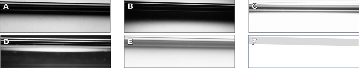
Same as Figure 8. A series of illusions created when a simple glass tube (tip of Pasteur pipette sloping down slightly to right) is viewed with light microscopy under varying conditions: (A) bright field compound scope @100×, substage condenser up; (B) same as A but condenser down; (C) same as A but stronger light; (D) same as A but phase contrast; (E) same tube but under a dissecting scope @40× and substage mirror at 45°; (F) is a drawing of the actual situation used to generate images A–E, with the gray bar in F representing the exact position of the compositional boundaries that generated A–E.
Current usage metrics show cumulative count of Article Views (full-text article views including HTML views, PDF and ePub downloads, according to the available data) and Abstracts Views on Vision4Press platform.
Data correspond to usage on the plateform after 2015. The current usage metrics is available 48-96 hours after online publication and is updated daily on week days.
Initial download of the metrics may take a while.