Figure 1.
Download original image
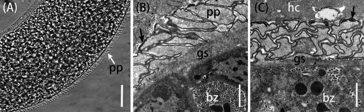
Morphological characteristics of Sarcocystis tenella sarcocysts from sheep muscle. (A) Light micrograph of a sarcocyst (unstained); note the sarcocyst wall with numerous palisade-like protrusions (pp). Scale bar = 20 μm. (B) Longitudinal section of sarcocyst wall by transmission electron microscopy (TEM). The sarcocyst wall had palisade-like protrusions (pp), characterized by the apex which contained dense plaques (arrow); a layer of ground substances (gs) located beneath the primary sarcocyst wall surrounded bradyzoites (bz). Scale bar = 1 μm. (C) Cross-section of a sarcocyst by TEM; note the dense plaques (arrow), ground substance (gs), bradyzoites (bz), and host cell (hc). Scale bar = 1 μm.
Current usage metrics show cumulative count of Article Views (full-text article views including HTML views, PDF and ePub downloads, according to the available data) and Abstracts Views on Vision4Press platform.
Data correspond to usage on the plateform after 2015. The current usage metrics is available 48-96 hours after online publication and is updated daily on week days.
Initial download of the metrics may take a while.