Figure 2.

Download original image
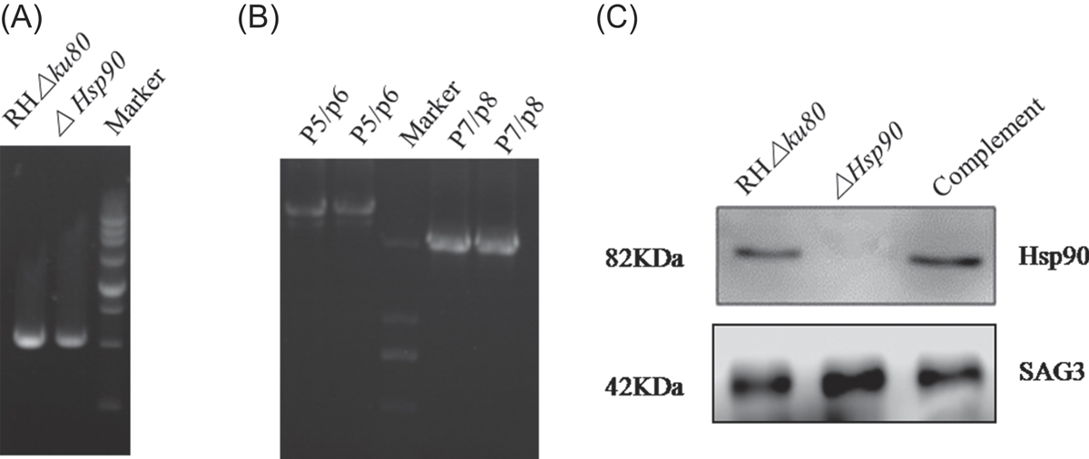
Confirmation of ΔHsp90 and complemented parasites. (A, B) PCR analysis of ΔHsp90 strains. The positions of the primers are shown in Figure 1A. P5/P6 and P7/P8 were used to amplify the conjunct regions of 5′ and 3′ integration of the Ble gene construct into the corresponding Hsp90 locus, respectively. (C) Western blotting analysis showing detection of Hsp90 in wild-type T. gondii RHΔku80 and in the complemented strain, but absence in ΔHsp90 parasites. Surface antigen (SAG3) was used as a loading control.
Current usage metrics show cumulative count of Article Views (full-text article views including HTML views, PDF and ePub downloads, according to the available data) and Abstracts Views on Vision4Press platform.
Data correspond to usage on the plateform after 2015. The current usage metrics is available 48-96 hours after online publication and is updated daily on week days.
Initial download of the metrics may take a while.


