Figures 1–6.
Download original image
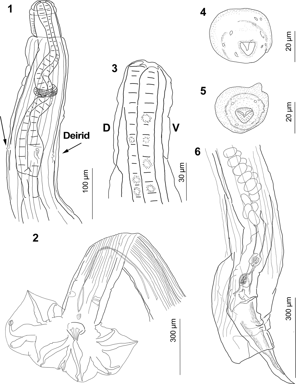
Guerrerostrongylus marginalis n. sp. 1, Ventral view of the anterior end of male, showing cephalic vesicle, esophagus, nerve ring, deirids (indicated by arrows), and excretory pore (between deirids). 2, Posterior end of a paratype, showing caudal bursa, genital cone, and spicules. 3, Lateral view of cephalic vesicle and stoma with esophageal tooth (upper left) not projected toward lumen. 4, Apical view of a female featuring dorsal tooth and triangular stoma. 5, Apical view of a male, showing dorsal tooth and triangular stoma. 6, Posterior end of a paratype showing cuticular invagination covering vulva, vulva, anus, ovejector, infundibulum, eggs in uterus, and tail.
Current usage metrics show cumulative count of Article Views (full-text article views including HTML views, PDF and ePub downloads, according to the available data) and Abstracts Views on Vision4Press platform.
Data correspond to usage on the plateform after 2015. The current usage metrics is available 48-96 hours after online publication and is updated daily on week days.
Initial download of the metrics may take a while.