Fig. 1.

Download original image
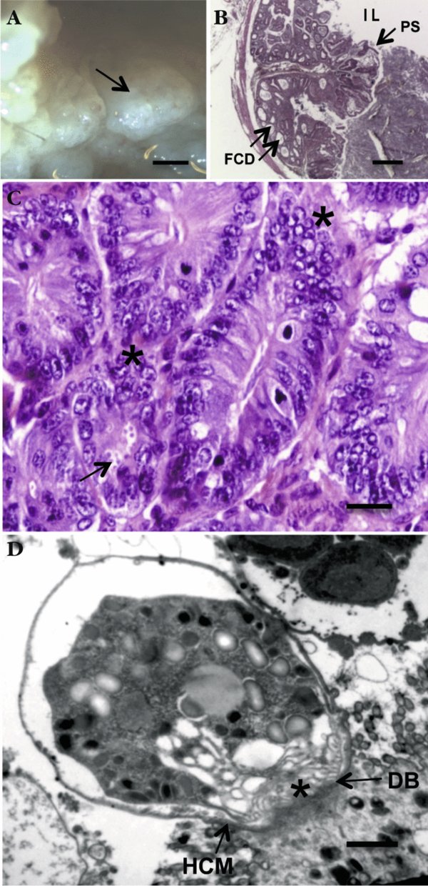
Ileocaecal adenocarcinoma induced by Cryptosporidium parvum in dexamethasone-treated SCID mice.
A: adenomatous masses (arrow) in the intestinal lumen. SCID mouse orally infected with C. parvum oocysts and euthanatized 45 days post-infection; B: projection of a polypoid structure (PS) with focal cystic dilations (FCD) developing inside the intestinal lumen (IL). Hematoxylin & Eosin staining; C: high grade epithelial neoplasia characterized by architectural distortion and cellular atypias (*): loss of normal polarity, nuclear stratification and prominent nucleoli, associated to the presence of numerous parasites inside the glands at the surface of epithelium (arrow). Dexamethasonetreated SCID mouse infected by C. parvum after 100 days postinfection. Hematoxylin & Eosin staining; D: transmission electron micrograph showing intracellular C. parvum developmental stages inside their parasitophorous vacuole. A feeder organelle (*) adheres to the host cell membrane (HCM) on an electron-dense area, the dense band (DB).
Bar (μm) = A: 1,000; B: 400; C: 25; D: 0.4.
Current usage metrics show cumulative count of Article Views (full-text article views including HTML views, PDF and ePub downloads, according to the available data) and Abstracts Views on Vision4Press platform.
Data correspond to usage on the plateform after 2015. The current usage metrics is available 48-96 hours after online publication and is updated daily on week days.
Initial download of the metrics may take a while.


