


| Issue |
Parasite
Volume 33, 2026
|
|
|---|---|---|
| Article Number | 38 | |
| Number of page(s) | 13 | |
| DOI | https://doi.org/10.1051/parasite/2026037 | |
| Published online | 09 July 2026 | |
Research Article
“Giant tick” attacks: dynamics of Hyalomma lusitanicum and detection of Rickettsia sibirica mongolitimonae in southern France
Attaques de « tiques géantes » : dynamique d’Hyalomma lusitanicum et détection de Rickettsia sibirica mongolitimonae dans le sud de la France
1
Aix Marseille Univ, SSA, RITMES, Marseille, France
2
IHU Méditerranée Infection, Marseille, France
3
Centre de référence (Sud) des maladies vectorielles liées aux tiques, AP-HM, Marseille, France
4
Unité Parasitologie Entomologie, Département Microbiologie et Maladies Infectieuses, Institut de Recherche Biomédicale des Armées, 13005 Marseille, France
5
Service Mer, Milieux Aquatiques et Zones Humides Direction, Pilotage du Grand Cycle de l'Eau, DGD Transition Environnementale, Eau, Culture et Sport, Aix Marseille Provence Métropole, Marseille, France
6
CIRAD, UMR ASTRE, Montpellier, France
7
ASTRE, Univ Montpellier, CIRAD, INRAE, Montpellier, France
8
Institut de recherche pour le développement (IRD), Maladies Infectieuses, Négligées et Émergentes au Sud (MINES), Marseille, France
* Corresponding author: This email address is being protected from spambots. You need JavaScript enabled to view it.
Received:
23
March
2026
Accepted:
17
June
2026
Abstract
Since 2019, media reports have raised concerns about the emergence of “giant ticks” in Europe, particularly Hyalomma marginatum, due to its potential to transmit the Crimean-Congo haemorrhagic fever virus (CCHFV). In 2022, following several reports of unusually large ticks displaying aggressive behaviour toward humans in the Étang de Bolmon area (Bouches-du-Rhône, southern France), a popular area for walking and nature exploration, an investigation was conducted to characterise the local tick fauna, their seasonal activity, and associated microorganisms. Between 2022 and 2025, a total of 2,940 ticks were collected through environmental and host-based sampling, with 89% obtained from the ground and 11% from rabbits. Morphological, molecular, and MALDI-TOF mass spectrometry analyses were carried out and four species were identified: Hyalomma lusitanicum (78%), Rhipicephalus pusillus (22%), Haemaphysalis hispanica (<1%), and Ixodes ventalloi (<1%). The “giant ticks” were identified as H. lusitanicum, not H. marginatum. Molecular screening revealed Rickettsia sibirica mongolitimonae in H. lusitanicum, suggesting its role in the ecology of this emerging human pathogen, along with the endosymbiont Candidatus Midichloria mitochondrii. This work also confirmed the accuracy of MALDI-TOF MS as a reliable tool for rapid tick identification and highlights the re-emergence of H. lusitanicum in southern France, while indicating that the associated risk to human public health currently remains relatively limited.
Résumé
Depuis 2019, les médias ont fait part de leur inquiétude quant à l’émergence de « tiques géantes » en Europe, notamment Hyalomma marginatum, en raison de son potentiel de transmission du virus de la fièvre hémorragique de Crimée-Congo (VFHCC). En 2022, suite à plusieurs signalements de tiques de taille inhabituelle présentant un comportement agressif envers l’homme dans la région de l’Étang de Bolmon (Bouches-du-Rhône, sud de la France), un site prisé pour la randonnée et la découverte de la nature, une étude a été menée afin de caractériser la faune locale de tiques, son activité saisonnière et les micro-organismes associés. Entre 2022 et 2025, 2 940 tiques ont été collectées par échantillonnage environnemental et sur leurs hôtes, dont 89 % prélevées au sol et 11 % sur des lapins. Les analyses morphologiques, moléculaires et par spectrométrie de masse MALDI-TOF ont permis d'identifier quatre espèces : Hyalomma lusitanicum (78 %), Rhipicephalus pusillus (22 %), Haemaphysalis hispanica (< 1 %) et Ixodes ventalloi (< 1 %). Les « tiques géantes » ont été identifiées comme H. lusitanicum, et non H. marginatum. Le criblage moléculaire a révélé la présence de Rickettsia sibirica mongolitimonae chez H. lusitanicum, suggérant son rôle dans l'écologie de ce pathogène humain émergent. L'endosymbiote Candidatus Midichloria mitochondrii a également été detecté. Ces travaux ont également confirmé la fiabilité de la spectrométrie de masse MALDI-TOF pour l'identification rapide des tiques et ont mis en évidence la réémergence de H. lusitanicum dans le sud de la France, tout en indiquant que le risque associé pour la santé publique reste actuellement relativement limité.
Key words: Ticks / Hyalomma lusitanicum / Rhipicephalus pusillus / Ixodes ventalloi / Haemaphysalis hispanica / Rickettsia sibirica mongolitimonae
Edited by Jean-Lou Justine.
© P. Parola et al., published by EDP Sciences, 2026
 This is an Open Access article distributed under the terms of the Creative Commons Attribution License (https://creativecommons.org/licenses/by/4.0), which permits unrestricted use, distribution, and reproduction in any medium, provided the original work is properly cited.
Introduction
Ticks are obligate, blood-feeding, temporary ectoparasitic arthropods belonging to the subclass Acari and the order Ixodida. They are divided into two main families: Ixodidae (hard ticks), comprising over 700 species worldwide, and Argasidae (soft ticks), comprising roughly 200 species [5]. Ticks feed on mammals, birds, and reptiles, and many species may also bite and feed on humans. Moreover, ticks can act as vectors by efficiently transmitting some of the microorganisms they carry. Importantly, they harbor a vast range of microorganisms, including bacteria, viruses, and protozoa, some of which are pathogenic to humans and other animals [23].
In Europe, the tick genera most frequently implicated in human infectious diseases are Ixodes, Rhipicephalus, and Dermacentor spp. [30]. In France, four tick species are currently considered of medical importance. Ixodes ricinus is the main vector of spirochetes belonging to the Borrelia burgdorferi sensu lato complex responsible for Lyme disease. It may also harbor several other tick-borne bacterial agents, including Rickettsia spp., and is a vector of tick-borne encephalitis virus in north-eastern France. Dermacentor marginatus, the ornate sheep tick, and Dermacentor reticulatus, the ornate dog tick, are recognized vectors of spotted fever group (SFG) rickettsioses. Rhipicephalus sanguineus sensu stricto, the brown dog tick, is known as the vector of Rickettsia conorii conorii, the causative agent of potentially life-threatening Mediterranean spotted fever in southern France. Rhipicephalus sanguineus is also known to harbor other rickettsial organisms, including human pathogens [35, 47, 50].
More recently, Hyalomma marginatum has attracted growing attention in France. Until recent years, continental France was considered free of this large tick species; however, the presence of reproducing populations of H. marginatum in parts of southern France has now been demonstrated [3]. This tick is known to harbor several human pathogens, including the Crimean-Congo hemorrhagic fever virus (CCHFV), and has therefore become a focus of increased investigation in France [1, 3, 4, 32].
Since 2019, several media outlets have raised public concern regarding the emergence and potential spread of “giant ticks” (H. marginatum) in Europe, including in France [11]. In 2022, as a national reference center for tick-borne diseases, we were alerted by local walkers to the presence of unusually large and aggressive ticks in a specific area of southern France (Étang de Bolmon, Bouches-du-Rhône) and decided to investigate the exact location where these encounters occurred.
The primary objective of this study was to collect and identify ticks in this area and to assess their annual dynamics. Secondary objectives included further assessing matrix-assisted laser desorption/ionization time-of-flight mass spectrometry (MALDI-TOF MS) for tick identification and detecting specific microorganisms carried by these ticks using molecular tools.
Material and methods
Ethics
Management of the site was transferred in 2017 to the Aix-Marseille Provence Métropole, the large intermunicipal authority (a “metropolis” in the French administrative system) that governs the wider Marseille region in southern France [13]. Investigations and research activities were conducted under the supervision of the Sea, Aquatic Environments and Wetlands Department within the Directorate for Integrated Water Cycle Management. Ferreting operations are authorized through an application that hunters must submit each year to the Coastal Protection Agency (Conservatoire du littoral), the Prefecture, and the Bouches-du-Rhône hunters’ federation. In the current work, these operations were supervised by a member of the Sea, Aquatic Environments and Wetlands Department (EG), and a veterinarian. The rabbits are subsequently released into other hillside sites (Côte Bleue) to support the repopulation of hunted areas and prevent the introduction of Spanish rabbit strains. The rabbits are released, free of arthropods to avoid arthropod expansion. Furthermore, under French legislation, collecting ticks in natural environments for research purposes is not subject to any specific authorization requirement. Public participatory research programs on ticks, coordinated by French research institutions, explicitly encourage citizens to collect and submit ticks for scientific research purposes, without reference to any prior authorization requirement [12].
Study area
Our study was conducted at Étang de Bolmon (Bolmon pond) [13, 14, 52]. This site covers 600 ha and comprises a mosaic of natural landscapes, including a low-salinity lagoon, temporary Mediterranean marshes, wet meadows, salt marshes, steppe grasslands, woodlands (pine and riparian forests), and a dune lido. It is located near the Salins du Lion natural complex and is surrounded by paths designed for walkers and cyclists. Étang de Bolmon is listed as a natural zone of ecological, faunal, and floral interest; it is classified as a sensitive natural area under Article L146-6 of the Coastal Law, registered in the National Wetland Inventory, and designated as a Natura 2000 site. More specifically, the protected natural site of Étang de Bolmon has been under the land protection of the Coastal protection Agency (Conservatoire du Littoral), which has owned it since 1992. The area includes a discovery trail that allows visitors to explore the local flora and fauna, with interpretive panels along the way. Étang de Bolmon is well known as an excellent site for walking and nature observation (Supplementary Figure 1). The area is used year-round, with increased frequentation during periods of favorable weather.
A nature protection and environmental education association specializing in entomology and active in this area alerted our teams to unusual “giant tick” attacks on walkers and picnickers. Investigations were therefore conducted in the specific area where these incidents were reported (43° 24′ 47″ N, 5° 10′ 41″ E).
Field capture of ticks
Tick sampling was conducted between July 2022 and September 2025, with a total of 24 field surveys performed during this period (Table 1). Sampling was carried out at variable frequencies until June 2024, followed by bi-monthly surveys until August 2024, and monthly surveys from September 2024 to September 2025.
Field collection of ticks at Étang de Bolmon, southern France (2022–2025) and their morphological identification. A timeline can be seen as Supplementary Figure 3.
Ticks were collected during field trips using several methods, including direct visual searches by collectors walking through the area to spot ticks on the ground, and the use of dry ice to attract actively host-seeking ticks through the release of CO2 during sublimation. During the first sampling sessions, flagging was also performed over random 10 m transects. This consisted of dragging a piece of fabric – such as a small blanket – across vegetation behind the observer, allowing questing ticks to attach to the cloth as it passed (Fig. 1) [39, 40]. As soon as ticks were observed, they were collected manually by team members searching for active ticks on the ground. The number of collectors per field session ranged from three to six, with each team collecting for 60–90 min. See supplementary videos illustrating tick collection at: https://doi.org/10.5281/zenodo.20141476.
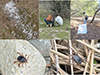 |
Figure 1 Field collection sites for ticks at Étang de Bolmon using flagging (A), dry ice placed on the ground to attract ticks (B), and direct visual searches by collectors (C). (D, E) Male Hyalomma lusitanicum ticks on the ground. See supplementary videos illustrating tick collection at: https://doi.org/10.5281/zenodo.20141476. |
Fieldwork also included participation in three ferreting operations in February and March 2023 – a traditional method of rabbit control. The goal of ferreting is to drive rabbits out of their burrows using trained ferrets (Mustela putorius), small and slender carnivorous mammals that enter the burrows and chase rabbits into nets placed at the exits. Ticks were collected from rabbits using tweezers (Supplementary Figure 2). Falcon tubes containing ticks were transported to the laboratory and stored at –20 °C after the ticks had been counted and morphologically identified. The rabbits were maintained by hunters as part of authorized hunting activities. Many fleas were also collected on this occasion (to be reported elsewhere).
Morphological and molecular identification of ticks
All collected ticks were first morphologically identified using reference documents [50], under a Leica EZ4 stereomicroscope (Leica Microsystems, Nanterre, France) and a Zeiss Axio Zoom V16 microscope (Zeiss, Marly-le-Roi, France). Ticks were sorted by species and sex subsequently stored at −20 °C until further analyses. A subset of specimens from each morphologically identified species was randomly selected for molecular confirmation by amplification of a 465 bp fragment of the tick 16S rRNA gene, as previously described [17].
Each selected tick was bisected longitudinally. Genomic DNA was extracted individually from tick halves and stored at −20 °C until use, as previously described [17, 46]. DNA samples were then subjected to standard PCR using an automated thermal cycler [17]. DNA extracted from R. sanguineus sensu stricto (temperate lineage) specimens from our laboratory colonies served as a positive control, while the PCR mix without template DNA was used as a negative control. Following visualization on agarose gel electrophoresis, positive PCR products were purified and sequenced as described previously [17, 37, 41]. The obtained sequences were assembled and analyzed using ChromasPro software (version 1.7.7; Technelysium Pty. Ltd., Tewantin, Australia) and subsequently compared with sequences available in GenBank using BLAST (http://blast.ncbi.nlm.nih.gov/).
Identification of ticks by MALDI-TOF MS
The four legs on the same side of each tick were removed, homogenized, and subjected to MALDI-TOF MS analysis with a Microflex LT mass spectrometer (Bruker Daltonics, Bremen, Germany), as previously described [35, 43, 60]. The quality and reproducibility of MS profiles obtained from tick legs – defined by the absence of background noise and peak intensities exceeding 3,000 arbitrary units – were visually assessed using FlexAnalysis v3.3 software (Bruker Daltonics, Bremen, Germany). Only spectra fulfilling these criteria were selected for further analysis. The reproducibility and species-specificity of MS spectra were evaluated using cluster analysis (MSP dendrogram). The dendrogram was generated by clustering specimens based on comparisons of their protein mass profiles (i.e., mass signals and intensities). Based on the consistency between morphological and molecular identification, MALDI-TOF MS spectra from reference specimens of each tick species were incorporated into our in-house arthropod MALDI-TOF MS reference spectra database. Blind tests were subsequently performed by matching the MS spectra of tick legs from remaining specimens against the updated reference database. The reliability of species identification was expressed by log score values (LSVs), ranging from 0 to 3, generated by MALDI-Biotyper v3.0 software. Each LSV corresponded to the degree of similarity between the MS spectra of tested samples and those of reference spectra in the database [35, 43, 60].
DNA extraction and microorganism screening
DNA was individually extracted from one half of each tick, randomly selected from ticks collected during each sampling period between July 5, 2022 and August 22, 2025, as described above. The extracted DNA was screened by quantitative PCR (qPCR) to detect Bartonella spp., Borrelia spp., Coxiella burnetii, members of the Anaplasmataceae family, and Rickettsia spp., using primers and probes previously described [16, 45, 46]. DNA from ticks collected between July 5, 2022 and August 8, 2024 was tested individually, whereas DNA from ticks collected between August 2024 and May 2025 was tested in pools of 4–5 specimens. This pooling strategy was implemented to manage the large number of collected ticks, reduce reagent costs, and accelerate testing without compromising detection efficiency. qPCR assays were performed under conditions identical to those previously described [66], and positive controls were included in all runs.
DNA extracted from reference strains (Rickettsia montanensis, Bartonella elizabethae, Anaplasma phagocytophilum, C. burnetii, and Borrelia crocidurae) served as specific positive controls. For pooled tick DNA samples, synthetic plasmid controls containing the corresponding target sequences were used. Plasmid aliquots were prepared at a concentration of 1.0 × 10⁵ copies/5 μL and included as positive controls in each assay [7, 18]. DNA extracted from R. sanguineus s.s. from our laboratory colonies (known to be free of the bacteria tested) served as a negative control. Samples were considered positive when the qPCR cycle threshold (Ct) value was below 36 [17].
Samples testing positive for Rickettsia spp. by qPCR were subsequently analyzed using a specific qPCR assay targeting a Rickettsia sibirica mongolitimonae gene fragment [57]. All R. sibirica mongolitimonae-positive samples were further subjected to conventional PCR and sequencing of the ompA (outer membrane protein A) and gltA (citrate synthase) genes, as previously described [24]. Samples that tested positive for members of the Anaplasmataceae family by qPCR, specifically those with Ct values <28, were analyzed by conventional PCR and sequencing of the Ehrlichia 16S rRNA gene, amplifying a 345 bp fragment specific to Anaplasmataceae bacteria [49].
Following amplification, PCR products were resolved by electrophoresis on 1.5% agarose gels stained with SYBR Safe™ (Thermo Fisher Scientific, Illkirch, France) and visualized using a ChemiDoc MP UV imager (Bio-Rad, Marnes-la-Coquette, France). PCR products were purified and sequenced using a BigDye Terminator kit (Thermo Fisher Scientific, Illkirch, France) on a Genetic Analyzer ABI PRISM 3130 (Applied Biosystems, Courtaboeuf, France). Sequence data were analyzed and assembled using ChromasPro software (version 1.34; Technelysium Pty Ltd., Tewantin, QLD, Australia) and compared with reference sequences available in GenBank using the BLAST tool, as previously described [17].
Results
Sampling collection and morphological identification of ticks
A total of 2,940 ticks were collected during 24 field outings at the Étang de Bolmon site, including two outings in 2022, four in 2023, nine in 2024, and nine in 2025 (Table 1; Supplementary Figure 3). Among these, 1,199/2,940 (40.8%) were collected during 12 summer outings (June–August), 350/2,940 (11.9%) during four autumn outings (September–November), 455/2,940 (15.5%) during three winter outings (December–February), and 925/2,940 (31.5%) during five spring outings (March–May) (Table 1). Of the total, 2,616/2,940 (89%) ticks were collected from the ground beneath trees during 21 field outings, whereas 324/2,940 (11%) were removed from rabbits during ferreting operations (Table 1). Among the 2,616 ground-collected ticks, 956 (36.5%) were obtained during nine collections conducted without dry ice, and 1,660 (63.5%) during 12 collections using dry ice. Overall, 2,928/2,940 (99.6%) of the collected ticks were adults, including 1,544/2,928 (52.7%) females. The remaining 12/2,940 (0.4%) were nymphs. Based on morphological characteristics, ticks were identified as belonging to four species: Hyalomma lusitanicum (n = 2,279), Rhipicephalus pusillus (n = 647), Haemaphysalis hispanica (n = 12), and Ixodes ventalloi (n = 2).
All H. lusitanicum specimens were collected from the ground beneath trees, except for one specimen collected during a ferreting operation, but found unattached on the ground. All H. hispanica were collected from rabbits during ferreting operations. The I. ventalloi nymph was collected from a rabbit, whereas the adult was collected from the ground. Lastly, R. pusillus specimens were collected both from rabbits and from the ground (Tables 1, Supplementary Table 1).
Molecular and MALDI-TOF MS identification of ticks
A total of 23 tick specimens, including 10 H. lusitanicum, 10 R. pusillus, two H. hispanica, and one I. ventalloi, were subjected to molecular identification to confirm morphological classification, using amplification and sequencing of the 16S rRNA gene. High-quality sequences were obtained for 20/23 samples (87%), including 10/10 (100%) H. lusitanicum, 7/10 (70%) R. pusillus, 2/2 (100%) H. hispanica, and 1/1 (100%) I. ventalloi. Intra-species comparison revealed 100% identity among H. lusitanicum sequences, 99.7–100% among R. pusillus, and 100% among H. hispanica. BLAST analysis showed that H. lusitanicum 16S rRNA gene sequences exhibited 98% query coverage and 99.75% identity with the H. lusitanicum reference sequence from Portugal (GenBank: KU130444), and 99.50 (GenBank: LC508315) – 99.74% (GenBank: ON366981) identity with other H. lusitanicum sequences in GenBank.
Additionally, R. pusillus sequences showed 100% query coverage and 99.77% identity with an R. pusillus sequence from Spain (GenBank: MZ420713). Haemaphysalis hispanica sequences exhibited 97% query coverage and 98.7% identity with a sequence obtained from Oryctolagus cuniculus in Spain (GenBank: MZ420715). The I. ventalloi sequence displayed 100% coverage and 100% identity with a sequence obtained from a bird in Italy (GenBank: MW175443).
Sequences obtained in this study from two H. lusitanicum, two R. pusillus, two H. hispanica, and one I. ventalloi have been deposited in GenBank under accession numbers PV577800–PV577801, PV577802–PV577803, PV919885–PV919886, and PV919887, respectively.
Legs from 422 H. lusitanicum, 349 R. pusillus, six H. hispanica, and one I. ventalloi were subjected to MALDI-TOF MS analysis. Mass spectra analyzed using FlexAnalysis v3.3 software showed high signal intensities (>3,000 a.u., with no background noise) in 96.4% (750/778) of tick analyses (Table 2). MS spectra visualization for one to five specimens per species demonstrated both intra-species reproducibility and inter-species specificity. These results were supported by MSP dendrograms, which showed that specimens of the same species consistently clustered on the same branch (Fig. 2).
Results of MALDI-TOF MS analysis of ticks collected at Étang de Bolmon, Southern France (2022–2025).
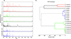 |
Figure 2 Comparison of MS spectra from four tick species collected at Étang de Bolmon (A) Representative MS spectra of Haemaphysalis hispanica (1–2), Hyalomma lusitanicum (3–4), Ixodes ventalloi (5), and Rhipicephalus pusillus (6–7). (B) Dendrogram constructed from the MS spectra of H. hispanica, H. lusitanicum, I. ventalloi, and R. pusillus using MALDI-Biotyper v3.3 software. |
Among the MS spectra corresponding to 20 specimens confirmed by molecular analysis, seven spectra of H. lusitanicum, six of R. pusillus, two of H. hispanica, and one of I. ventalloi were selected and added to our in-house MALDI-TOF MS reference database. The remaining 734 MS spectra – comprising 397 from H. lusitanicum, 333 from R. pusillus, and four from H. hispanica based on morphological identification – were blindly tested against the updated database. All 397 H. lusitanicum specimens were correctly identified, with log score values (LSVs) ranging from 1.70 to 2.49 (mean ± SD: 2.03 ± 0.15), and 374/397 (94.2%) achieving LSVs ≥1.8. In addition, R. pusillus specimens were correctly identified with LSVs between 1.71 and 2.74 (mean ± SD: 1.95 ± 0.10), and 289/333 (86.8%) achieving LSVs ≥1.8. Lastly, H. hispanica specimens were also correctly identified, with LSVs ranging from 1.76 to 2.03 (mean ± SD: 1.88 ± 0.07), and 3/4 (75%) achieving LSVs ≥1.8. Raw MALDI-TOF MS spectra of H. lusitanicum, R. pusillus, H. hispanica, and I. ventalloi are available at: https://doi.org/10.5281/zenodo.20187728.
Screening for specific bacteria
DNA extracts from 1,360 H. lusitanicum, 454 R. pusillus, six H. hispanica, and one I. ventalloi were tested by qPCR either individually or in pools for the presence of microorganisms. In total, 1,062 H. lusitanicum and 222 R. pusillus specimens were combined into 214 and 45 pools, respectively.
All ticks, whether tested individually or in pools, were negative for Borrelia spp., Bartonella spp., and Coxiella burnetii. Two pools of H. lusitanicum, collected in September 2024 and February 2025, tested positive for Rickettsia spp., with Ct values of 34 and 35. Both pools were positive for R. sibirica mongolitimonae by species-specific qPCR (Ct = 31 and 34). Individual testing of the ten specimens comprising these two pools revealed four positives (Ct = 29, 32, 35, and 36). Standard PCR and sequencing of these four specimens using the ompA and gltA genes yielded two usable ompA sequences and one usable gltA sequence. BLAST analysis showed 100% coverage and 99.81% identity for ompA (GenBank: MF379309.1) and 100% coverage with 99.08% identity for gltA (GenBank: DQ423370.1). The three R. sibirica mongolitimonae sequences from this study were deposited in GenBank under accession numbers PX580606–PX580608.
A total of 111 pools of H. lusitanicum and 12 pools of R. pusillus tested positive for bacteria belonging to the family Anaplasmataceae, with positive H. lusitanicum pools detected during all sampling periods. Thirty H. lusitanicum specimens, grouped into six pools with Ct < 28, were selected for conventional PCR and sequencing of the Anaplasmataceae 16S rRNA gene. Usable sequences were obtained from six specimens. BLAST analysis revealed 100% coverage and 99.62–100% identity with Candidatus M. mitochondrii (GenBank: KX359182.1). These six Candidatus M. mitochondrii sequences were deposited in GenBank under accession numbers PX417374–PX417379. The 12 DNA pools of R. pusillus that tested positive for Anaplasmataceae had Ct values >28 and were therefore not selected for conventional PCR and sequencing.
Discussion
In this study, we identified several tick species, including those responsible for the so-called “giant tick” attacks in a specific area of southern France, Étang de Bolmon, a popular location for walking and exploring nature. The sampling effort varied over time in both frequency and methodology, and multiple collection techniques were used. This heterogeneity in field methods likely introduced some bias in species representation and life-stage distribution. However, despite these limitations, our study yielded unique results, addressing citizen reports of attacks by unusually large ticks that generated media attention, as well as enabling the rediscovery of H. lusitanicum and the characterization of microorganisms associated with ticks from the Étang de Bolmon area.
Tick identification was initially performed using morphological criteria. Although unambiguous diagnostic features were observed in both males and females, species identification was definitively confirmed using molecular tools, which have been routinely employed in entomological research for the past two decades [71]. We also used MALDI-TOF MS, a proteomic tool that has recently emerged in the field of entomology, providing an opportunity to further assess its robustness for arthropod identification. Initially developed for clinical laboratories, MALDI-TOF MS was later applied to the diagnosis of genetic diseases and microorganisms, and it has recently gained prominence in entomology [65].
Previous studies by our group have detailed this approach for identifying both field-collected and laboratory-reared arthropods. LSVs greater than 1.8 are considered reliable for species identification, according to previous studies [43, 65]. Despite its advantages, MALDI-TOF MS does have limitations. Accurate identification requires comparison with reference spectra from specimens definitively identified by morphological or molecular methods. Preservation techniques can also influence protein spectra, and spectral variations may occur depending on the geographic origin of specimens [65]. Therefore, maintaining an up-to-date reference database by regularly adding new specimens is essential to ensure accurate identifications [65]. The present work provides further evidence of the reliability of MALDI-TOF MS, which was evaluated on a subset of the collected ticks.
In this study, the “giant ticks” were unexpectedly identified as H. lusitanicum rather than H. marginatum, contrary to initial assumptions based on the known distribution of tick species in southern France along the Mediterranean coast, as well as recent media coverage and public health concerns regarding H. marginatum and its potential role in transmitting the Crimean-Congo hemorrhagic fever virus [3].
Hyalomma lusitanicum are indeed large ticks, as unfed males and females measure 4–6 mm in length and possess a long capitulum (Fig. 3). They also actively host-seek and are attracted by carbon dioxide emitted through host respiration and other odors, which explains the effectiveness of dry ice traps in their capture [48]. The distribution of H. lusitanicum is typically restricted to the xeric or warm scrublands of the western Mediterranean basin (Portugal, Spain, Sicily, North Africa) [63]. The species is adapted to mesomediterranean vegetation and is frequently found within rock crevices, vegetal litter, or rabbit burrows. Also, H. lusitanicum is a three-host tick with selective parasitic behavior: its distribution is closely linked to that of the wild rabbit (O. cuniculus), on which immatures feed, while adults mainly infest ungulates and occasionally carnivores. Its immature stages develop in rabbit burrows, where microclimatic conditions are favorable. However, immatures may also feed on birds. Consequently, the tick may be abundant in areas hosting both rabbits and ungulates, such as cattle, wild boar, and deer [53, 69]. Although more regular sampling would have been required, our data suggest that the peak of adult activity occurs in late spring/early summer, which is consistent with the literature. Importantly, larvae are known to be active from May to September, nymphs from July to September, and adults throughout the year, with peaks in May–July and October–November [2, 42, 68, 69]. Notably, in our study, ticks were also collected during winter.
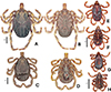 |
Figure 3 Representative images of Hyalomma lusitanicum (A, female dorsal view; B, female ventral view; C, male dorsal view; D, male ventral view) and Rhipicephalus pusillus (E, female dorsal view; F, female ventral view; G, male dorsal view; H, male ventral view) specimens collected in this study conducted at Étang de Bolmon, Southern France. Scale bar: 1 mm. |
In France, H. lusitanicum can be considered an emerging or re-emerging species. It has been reported sporadically in the southernmost regions, particularly in saline and alluvial habitats of the Rhône delta [50]. The species was collected until the late 1950s in Camargue, Pyrénées-Orientales, and the Var [42, 55]. Its apparent disappearance after the 1950s was attributed to myxomatosis, which decimated 90–98% of wild rabbits in France between 1952 and 1955 [25]. Later studies failed to recover H. lusitanicum in these areas [28, 56]. Gilot (1985) [26] suggested that the species might reappear episodically in small, localized hotspots following introductions. Given that European rabbits have been reintroduced from Spain over the past five decades [51], reintroduction of H. lusitanicum through parasitized hosts is plausible, though difficult to confirm. Similarly, although H. lusitanicum can be found on birds, their role in the dispersal of this species remains unclear [53, 69].
The Étang de Bolmon area offers suitable habitats for H. lusitanicum, although it primarily serves as a wintering site for migratory birds, which are unlikely hosts for this tick [2]. The site hosts large rabbit populations, regulated by controlled hunting, as well as occasional grazing cattle and horses. Despite its limited forest cover and ecological isolation, the site supports H. lusitanicum persistence. This study provides the most extensive investigation of the species’ presence in France, following sentinel alerts.
Other tick species identified, including R. pusillus, H. hispanica, and I. ventalloi, are rabbit-associated pholeophilous ticks adapted to burrow microhabitats. For example, R. pusillus, the second most prevalent species, is endophilic and monotropic, primarily infesting O. cuniculus [50]. It belongs to the R. sanguineus group and has recently been redescribed [22]. While typically restricted to rabbit burrows, R. pusillus can infest carnivores, domestic animals, and ungulates. Our observations of questing adults far from burrows confirm its occasional exophilous behavior, as previously noted [28].
Haemaphysalis hispanica, collected only during ferreting, is a small, rabbit-specific tick confined to the Mediterranean zone. After its first reports in France [42, 55], it was not detected again until the work of Gilot and colleagues [28, 50]. Ixodes ventalloi, also collected in this study, shares a similar ecology, being primarily associated with rabbit burrows but capable of parasitizing small mammals and birds [28].
From a public health perspective, tick-human interactions and potential pathogen transmission are key concerns. However, the mere fact of carrying a pathogen does not necessarily imply vectorial competence or the ability to transmit it.
Hyalomma lusitanicum can definitely bite humans, and adults exhibit active host-seeking (attacking) behavior. After the first field outings, we discontinued flagging collections because ticks were actively attacking or already visible on the ground. Regarding associated microorganisms, this tick species may be involved in the transmission of Coxiella burnetii to humans, the causative agent of Q fever [21]. Experimental models have suggested that this species is capable of maintaining the infection throughout its developmental cycle [34]. Among intracellular bacteria, two emerging human pathogens, Anaplasma phagocytophilum and Rickettsia aeschlimannii, have also been detected in H. lusitanicum. Both are known to be transmitted through tick bites [10, 68]. The presence of Francisella tularensis, the etiological agent of tularemia, as well as related endosymbionts (Francisella-like endosymbionts), has also been reported in this species [68]. Moreover, C. burnetii DNA was detected in wild lagomorphs in Spain, as well as in ticks collected from these animals, including H. hispanica, R. pusillus, and H. lusitanicum [6].
In a recent survey conducted in southern Spain to identify tick species parasitizing wild lagomorphs, several rickettsial pathogens (including Rickettsia massiliae, R. sibirica mongolitimonae, R. slovaca, and R. africae) were detected in H. lusitanicum ticks [58]. Several viruses have also been detected in this species, including the Crimean-Congo hemorrhagic fever virus (CCHFV), hepatitis E virus (HEV), and myxoma virus [68]. Regarding protozoa, the transmission capacity of Theileria annulata has long been suggested [33, 70], while Theileria equi and Babesia pecorum have also been detected, further emphasizing the veterinary importance of this tick [38, 68].
In the present study, we detected R. sibirica mongolitimonae in several H. lusitanicum specimens. This finding is particularly relevant for deciphering the ecology of this emerging human pathogen. Rickettsia sibirica mongolitimonae is a spotted fever group (SFG) Rickettsia first isolated from H. asiaticum ticks collected in Inner Mongolia, and later recognized as a human pathogen in France in 1996 [47]. According to a recent review by Santibáñez et al. (2025) [61], a total of 94 human cases have been reported to date, including 89 in Europe (mainly in Spain (n = 47), France (n = 36), Portugal, Greece, Türkiye, and North Macedonia), 4 in Africa, and 1 in Asia. Most cases occurred between April and July or in September. Clinical features include an inoculation eschar (sometimes multiple) at the tick bite site, a hallmark of many tick-borne SFG rickettsioses. Fever occurs in all published cases, and a generalized maculopapular rash was observed in nearly half of patients. Because a typical rope-like lymphangitis was reported in about 40% of cases, R. sibirica mongolitimonae infection has been referred to as lymphangitis-associated rickettsiosis.
The vector(s) of R. sibirica mongolitimonae have not been definitively confirmed. In Europe, this rickettsia has been detected in H. excavatum (Greece, Cyprus), H. marginatum (Spain), R. pusillus (Portugal, Spain, France), and Rhipicephalus bursa (Spain). In Turkey, it has been found in R. bursa, Haemaphysalis parva, H. excavatum, and H. marginatum. Among published human cases, four patients retained the tick responsible (one per patient), identified as female H. marginatum, H. excavatum, Hyalomma sp., and R. pusillus. Interestingly, one of the cases we previously reported occurred near Étang de Bolmon [67]. Another patient lived in Les Pennes-Mirabeau, an area of southern France where we have recently collected H. lusitanicum [57] (Stachurski et al. unpublished data).
In this study, we also detected Candidatus M. mitochondrii in H. lusitanicum ticks. This bacterium is an intracellular endosymbiont residing within the mitochondria of ovarian cells. It has been reported in numerous tick species worldwide, including Ixodes holocyclus, Amblyomma americanum, Rhipicephalus sanguineus, H. lusitanicum, and Rhipicephalus microplus [54]. It has been suggested that the abundance of “Ca. M. mitochondrii” in ticks may interfere with the detection of other pathogens [29].
Concerning R. pusillus, little is known about its affinity for biting humans. Based on years of field experience and an experimental attempt, Gilot reported that adult R. pusillus can bite and attach to humans, though attachment appears to be short-lived; no data exist for immature stages [27]. In our own investigations, we studied several tick-borne diseases in a household where R. pusillus was likely introduced by the family’s cat [19]. In the present work, we found evidence suggesting that R. pusillus can also bite humans, as some specimens were found on our clothing and footwear during fieldwork.
The role of R. pusillus as a vector of microorganisms remains poorly understood [64]. Among bacteria, Ehrlichia spp. and R. massiliae have been detected, the latter being recognized as an emerging zoonotic agent [58, 59]. Portuguese studies have reported the detection of R. sibirica mongolitimonae DNA in R. pusillus collected from a dead African mongoose (Herpestes ichneumon), alongside a serologically confirmed human case of infection in Portugal [15]. In 2013, we reported a cluster of one confirmed and two probable cases of lymphangitis-associated rickettsiosis caused by R. sibirica mongolitimonae in southern France, linked to a cat and R. pusillus ticks [19]. In the wild lagomorph study cited above, C. burnetii DNA was also detected in R. pusillus [6].
Finally, little is known about the two other tick species collected in this study. Interestingly, Gilot was unable to feed H. hispanica on humans [27]. In the recent molecular studies cited above, R. sibirica mongolitimonae and C. burnetii were also detected in H. hispanica [6, 58]. Regarding the affinity of I. ventalloi for biting humans, Gilot reported the first two records. According to him, due to its host specificity and pholeophilous behavior, human bites are likely rare. These ticks may come into contact with humans in two main ways: either through a hunted rabbit, or via another animal, e.g., domestic (e.g., a cat) or synanthropic (e.g., a hedgehog), that has attacked a rabbit or visited its burrow. In I. ventalloi, several microorganisms have been identified, including the Eyach virus [9], believed to be neuropathogenic to humans [8], and Anaplasma phagocytophilum, detected by molecular methods [62].
Conclusion
People visiting the Bolmon pond area should take protective measures against tick bites, including the use of protective clothing, repellents, insecticide-treated clothing and post-exposure tick checks [20]. The presence of ticks in the area could be indicated so that hikers are encouraged to check themselves for ticks at the end of their walk.
The drivers of H. lusitanicum introduction, establishment, and occasional disappearance in southern France are poorly understood. In addition to its documented association with rabbits, ecological factors such as temperature, vegetation structure (e.g., tree and plant composition), and soil characteristics, all of which are likely to play an important role in the ecology of Hyalomma spp., warrant further investigation. The role of birds also deserves to be studied.
Despite its recent occurrence, its presence remains discreet except in certain specific areas, such as the Étang de Bolmon. Interestingly, the term “giant ticks,” frequently used in the media to describe these ticks, is not a scientific designation, but rather a journalistic label intended to attract attention. Other tick species, including vector-competent ones such as Dermacentor spp., are also relatively large.
Regarding R. pusillus, since its anthropophily appears very limited – even among highly exposed individuals such as hunters or their associates handling heavily parasitized rabbits – its role as a potential vector of human diseases is likely low.
In this work, the detection of R. sibirica mongolitimonae in H. lusitanicum represents another step toward deciphering the ecology of this emerging human pathogen, which has most frequently been associated with Hyalomma spp. ticks and, less commonly, with R. pusillus. It should be noted, however, that the number of positive ticks was relatively low. An arthropod may harbor endosymbionts that are not transmitted, or may transiently carry microorganisms acquired through mixed infestations or bacteremic blood meals. The vector competence of H. lusitanicum remains to be definitively demonstrated, as does its potential public health importance.
According to our findings, the potential role of rabbits as reservoirs of this rickettsia warrants further investigation. Experimental feeding of infected ticks on laboratory animals should be attempted to assess whether the ticks can act as reservoirs for R. sibirica mongolitimonae through transovarial and transstadial transmission. To date, basic questions regarding the relationship between R. sibirica mongolitimonae and its tick vector(s) remain unresolved, and its life cycle, like those of many spotted fever group (SFG) rickettsioses, is still incompletely understood.
As a reference center for tick-borne and rickettsial diseases, we first focused on the detection of bacteria within our area of expertise. The results of our qPCR analyses did not yet allow us to fully address all questions regarding the public health relevance of these ticks, and further work is required. In recent years, metagenomic approaches have been increasingly used to study microorganisms carried by ticks [36, 44], including H. marginatum [31]. The next steps will be to investigate the virome and microbiome repertoires of H. lusitanicum and R. pusillus from Étang de Bolmon using metagenomic strategies. Consistent with previous reports, the high abundance of an endosymbiont reported here may mask less abundant pathogens in molecular analyses, highlighting the importance of deep metagenomic approaches to accurately assess tick-borne pathogen diversity.
Acknowledgments
We thank all the hunters who granted us access to the rabbits captured during ferreting operations.
Funding
This work was supported by a grant from the French Government managed by the National Research Agency under the “Investissements d’avenir (Investments for the Future)” program, with the reference ANR-10-IAHU-03 (Méditerranée Infection), by the Contrat Plan Etat-Région and European funding FEDER IHUPERF.
Author contribution statement
Conceived and designed the experiments: PP, LA, DZA, FS, EL. Performed the experiments: BO, BH, NM, ZA, AZD. Analyzed the data: BO, BH, ZA, AZD, LA. Contributed reagents/materials/analysis tools: ZA, AZD, LA, PP. Field collections preparation: ZA, AZD, BO, BH, LA, EL, NM, RB, PP, JMB, PP, FS. Drafted the paper: ZA, AZD, LA, PP. Investigation: ZA, AZD, BO, BH, LA, NM, RB, PP, JMB, PP, FS. Revised the paper critically: all authors.
Conflicts of interest
All authors have no conflicts of interest.
Supplementary material
Supplementary Table 1: Detail of ticks collection on rabbits at Étang de Bolmon, southern France, during ferreting operations, 2023. ♂ Male / ♀ Female. Access Supplementary Material
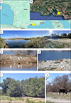 |
Supplementary Figure 1: Location and landscape of the Étang de Bolmon, Southern France yellow star in A and B), showing the pond (C ), birds (D), rabbits (E), picknickers (F), and cows (G). |
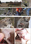 |
Supplementary Figure 2: Tick collection on European rabbits during ferreting operations at Étang de Bolmon, 2023, Southern France. |
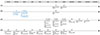 |
Supplementary Figure 3: Timeline of tick collection at Étang de Bolmon, Southern France, 2022 - 2025. Blue characters: collection during ferreting operations. |
References
- ANSES. 2023. Analyse des risques pour la santé humaine et animale liés aux tiques du genre Hyalomma en France. Avis de l'Anses. Rapport d'expertise collective. Available online: https://www.anses.fr/fr/system/files/SABA2020SA0039Ra.pdf. [Google Scholar]
- Apanaskevich DA, Santos-Silva MM, Horak IG. 2008. The genus Hyalomma Koch, 1844. IV. Redescription of all parasitic stages of H. (Euhyalomma) lusitanicum Koch, 1844 and the adults of H. (E.) franchinii Tonelli Rondelli, 1932 (acari: ixodidae) with a first description of its immature stages. Folia Parasitologica, 55, 61–74. [Google Scholar]
- Bah MT, Grosbois V, Stachurski F, Muñoz F, Duhayon M, Rakotoarivony I, Appelgren A, Calloix C, Noguera L, Mouillaud T, Andary C, Lancelot R, Huber K, Garros C, Leblond A, Vial L. 2022. The Crimean-Congo haemorrhagic fever tick vector Hyalomma marginatum in the south of France: modelling its distribution and determination of factors influencing its establishment in a newly invaded area. Transboundary and Emerging Diseases, 69, e2351–e2365. [Google Scholar]
- Bernard C, Joly Kukla C, Rakotoarivony I, Duhayon M, Stachurski F, Huber K, Giupponi C, Zortman I, Holzmuller P, Pollet T, Jeanneau M, Mercey A, Vachiery N, Lefrançois T, Garros C, Michaud V, Comtet L, Despois L, Pourquier P, Picard C, Journeaux A, Thomas D, Godard S, Moissonnier E, Mely S, Sega M, Pannetier D, Baize S, Vial L. 2024. Detection of Crimean–Congo haemorrhagic fever virus in Hyalomma marginatum ticks, southern France, May 2022 and April 2023. Eurosurveillance, 29, 2400023. [Google Scholar]
- Camicas J-L, Ed. 1998. Les Tiques du monde (Acarida, Ixodida): Nomenclature, stades décrits, hôtes, répartition (espèces décrites avant le 1/01/96) = The ticks of the world (Acarida, Ixodida): nomenclature, described stages, hosts, distribution (including new species described before 1/01/96). Paris: ORSTOM. p. 233, ISBN 2-7099-1418-2. [Google Scholar]
- Castro-Scholten S, Caballero-Gómez J, Martínez R, Nadales-Martín BJ, Cano-Terriza D, Jiménez-Martín D, Remesar S, Jiménez-Ruiz S, Gómez-Guillamón F, García-Bocanegra I. 2024. Occurrence of Coxiella burnetii in wild lagomorphs and their ticks in Spanish Mediterranean ecosystems. Zoonoses and Public Health, 71, 549–559. [Google Scholar]
- Charrel RN, La Scola B, Raoult D. 2004. Multi-pathogens sequence containing plasmids as positive controls for universal detection of potential agents of bioterrorism. BMC Microbiology, 4, 21. [Google Scholar]
- Chastel C. 1999. Erve et Eyach : deux virus isolés en France, neuropathogènes pour l’homme et largement répandus en Europe occidentale. Bulletin de l’Académie Nationale de Médecine, 182, 801–809. [Google Scholar]
- Chastel C, Main AJ, Couatarmanach A, Le Lay G, Knudson DL, Quillien MC, Beaucournu JC. 1984. Isolation of Eyach virus (Reoviridae, Colorado tick fever group) from Ixodes ricinus and I. ventalloi ticks in France. Archives of Virology, 82, 161–171. [Google Scholar]
- Chisu V, Zobba R, Foxi C, Pisu D, Masala G, Alberti A. 2016. Molecular detection and groEL typing of Rickettsia aeschlimannii in Sardinian ticks. Parasitology Research, 115, 3323–3328. [Google Scholar]
- Citique. 2025. Hyalomma marginatum – la tique géante !. Available online: https://www.citique.fr/Hyalomma-marginatum-la-tique-geante/. [Google Scholar]
- Citique. 2026. Citique | Des citoyens et des tiques – un programme de science participative ! Available online: https://www.citique.fr. [Google Scholar]
- Conservatoire du littoral. 2020. Convention de gestion du domaine terrestre et maritime du Conservatoire du littoral : site de l’Étang de Bolmon. Available online: https://deliberations.ampmetropole.fr/documents/metropole/deliberations/2020/12/17/rapportdelacommission/c0cxq.pdf. [Google Scholar]
- Conservatoire du littoral. 2025. Étang de Bolmon. Available online: https://www.conservatoire-du-littoral.fr/siteLittoral/246/28-etang-de-bolmon-13_bouches-du-rhone.htm. [Google Scholar]
- De Sousa R, Barata C, Vitorino L, Santos-Silva M, Carrapato C, Torgal J, Walker D, Bacellar F. 2006. Rickettsia sibirica isolation from a patient and detection in ticks, Portugal. Emerging Infectious Diseases, 12, 1103–1108. [Google Scholar]
- Diarra AZ, Almeras L, Laroche M, Berenger J-M, Koné AK, Bocoum Z, Dabo A, Doumbo O, Raoult D, Parola P. 2017. Molecular and MALDI-TOF identification of ticks and tick-associated bacteria in Mali. PLoS Neglected Tropical Diseases, 11, e0005762. [Google Scholar]
- Diarra AZ, Kone AK, Niare SD, Laroche M, Diatta G, Atteynine SA, Coulibaly M, Sangare AK, Kouriba B, Djimde A. 2020. Molecular detection of microorganisms associated with small mammals and their ectoparasites in Mali. American Journal of Tropical Medicine and Hygiene, 103, 2542–2551. [Google Scholar]
- Dubourg G, Edouard S, Prudent E, Fournier P-E, Raoult D. 2015. Incidental syphilis diagnosed by real-time PCR screening of urine samples. Journal of Clinical Microbiology, 53, 3707–3708. [Google Scholar]
- Edouard S, Parola P, Socolovschi C, Davoust B, La Scola B, Raoult D. 2013. Clustered cases of Rickettsia sibirica mongolitimonae infection, France. Emerging Infectious Diseases, 19, 337–338. [Google Scholar]
- Eisen L. 2022. Personal protection measures to prevent tick bites in the United States: knowledge gaps, challenges, and opportunities. Ticks and Tick-Borne Diseases, 13, 101944. [Google Scholar]
- Eldin C, Mélenotte C, Mediannikov O, Ghigo E, Million M, Edouard S, Mege J-L, Maurin M, Raoult D. 2017. From Q Fever to Coxiella burnetii infection: a paradigm change. Clinical Microbiology Reviews, 30, 115–190. [Google Scholar]
- Estrada-Peña A, Venzal JM, Nava S. 2018. Redescription, molecular features, and neotype deposition of Rhipicephalus pusillus Gil Collado and Ixodes ventalloi Gil Collado (Acari, Ixodidae). Zootaxa, 4446(3), 401–426. [Google Scholar]
- Estrada-Peña A. 2015. Ticks as vectors: taxonomy, biology and ecology. Revue Scientifique et Technique, 34, 53–65. [Google Scholar]
- Fournier P-E, Gouriet F, Brouqui P, Lucht F, Raoult D. 2005. Lymphangitis-associated rickettsiosis, a new rickettsiosis caused by Rickettsia sibirica mongolotimonae: seven new cases and review of the literature. Clinical Infectious Diseases, 40, 1435–1444. [Google Scholar]
- Giban J. 1956. Répercussion de la myxomatose sur les populations de lapin de garenne en France. Revue d’Écologie (La Terre et la Vie), 10(3–4), 179–188. [Google Scholar]
- Gilot B. 1985. Bases biologiques, écologiques et cartographiques pour l’étude des maladies transmises par les tiques (Ixodidae et Argasidae) dans les alpes françaises et leur avant-pays. Thèse doctorale, Université Scientifique et Médicale de Grenoble, Grenoble, France. [Google Scholar]
- Gilot B, Marjolet M. 1982. Contribution à l’étude du parasitisme humain par les tiques (Ixodidae et Argasidae), plus particulièrement dans le sud-est de la France. Médecine et Maladies Infectieuses, 12, 340–351. [Google Scholar]
- Gilot B, Rogers P, Lachet B. 1985. Données biologiques et écologiques sur les tiques de lagomorphes (et plus spécialement celles du lapin de garenne, Oryctolagus cuniculus L.) dans les alpes françaises et leur avant-pays. Acarologia, 26, 335–354. [Google Scholar]
- Gofton AW, Oskam CL, Lo N, Beninati T, Wei H, McCarl V, Murray DC, Paparini A, Greay TL, Holmes AJ, Bunce M, Ryan U, Irwin P. 2015. Inhibition of the endosymbiont “Candidatus Midichloria mitochondrii” during 16S rRNA gene profiling reveals potential pathogens in Ixodes ticks from Australia. Parasites & Vectors, 8, 345. [Google Scholar]
- Heyman P, Cochez C, Hofhuis A, Van Der Giessen J, Sprong H, Porter SR, Losson B, Saegerman C, Donoso-Mantke O, Niedrig M, Papa A. 2010. A clear and present danger: tick-borne diseases in Europe. Expert Review of Anti-Infective Therapy, 8, 33–50. [CrossRef] [PubMed] [Google Scholar]
- Joly-Kukla C, Bernard C, Bru D, Galon C, Giupponi C, Huber K, Jourdan-Pineau H, Malandrin L, Rakotoarivony I, Riggi C, Vial L, Moutailler S, Pollet T. 2024. Spatial patterns of Hyalomma marginatum-borne pathogens in the Occitanie region (France), a focus on the intriguing dynamics of Rickettsia aeschlimannii. Microbiology Spectrum, 12, e01256-24. [Google Scholar]
- Joly-Kukla C, Stachurski F, Duhayon M, Galon C, Moutailler S, Pollet T. 2024. Temporal dynamics of the Hyalomma marginatum-borne pathogens in southern France. Current Research in Parasitology & Vector-Borne Diseases, 6, 100213. [Google Scholar]
- Jongejan F, Uilenberg G. 2004. The global importance of ticks. Parasitology, 129, S3–S14. [CrossRef] [PubMed] [Google Scholar]
- González J, González MG, Valcárcel F, Sánchez M, Martín-Hernández R, Tercero JM, Olmeda AS. 2020. Transstadial transmission from nymph to adult of Coxiella burnetiid by naturally infected Hyalomma lusitanicum. Pathogens, 9(11), 884. [Google Scholar]
- Jumpertz M, Sevestre J, Luciani L, Houhamdi L, Fournier P-E, Parola P. 2023. Bacterial agents detected in 418 ticks removed from humans during 2014–2021, France. Emerging Infectious Diseases, 29, 701–710. [Google Scholar]
- Kipp EJ, Lindsey LL, Khoo B, Faulk C, Oliver JD, Larsen PA. 2023. Metagenomic surveillance for bacterial tick-borne pathogens using nanopore adaptive sampling. Scientific Reports, 13, 10991. [Google Scholar]
- Kumsa B, Laroche M, Almeras L, Mediannikov O, Raoult D, Parola P. 2016. Morphological, molecular and MALDI-TOF mass spectrometry identification of ixodid tick species collected in Oromia, Ethiopia. Parasitology Research, 115, 4199–4210. [Google Scholar]
- Luciano A, Jallow BJ, Liu M, Ma Y, Miambo RD, Meng F. 2025. Distribution of Rhipicephalus microplus and Hyalomma lusitanicum, and the pathogens they are carrying: A systematic review. Parasite Epidemiology and Control, 30, e00437. [Google Scholar]
- Mays Maestas SE, Maestas LP, Kaufman PE. 2025. Pathogen and host associations of soft ticks collected in South Texas. Vector-Borne and Zoonotic Diseases, 25, 34–42. [Google Scholar]
- Mays SE, Houston A, Trout Fryxell R. 2016. Comparison of novel and conventional methods of trapping ixodid ticks in the southeastern USA. Medical and Veterinary Entomology, 30, 123–134. [Google Scholar]
- M’madi SA, Diarra AZ, Almeras L, Parola P. 2022. Identification of ticks from an old collection by MALDI-TOF MS. Journal of Proteomics, 264, 104623. [Google Scholar]
- Morel P-C. 1959. Les Hyalomma (Acariens, Ixodidae) de France. Annales de Parasitologie Humaine et Comparée, 34, 552–555. [Google Scholar]
- Ngnindji-Youdje Y, Diarra AZ, Lontsi-Demano M, Berenger J-M, Tchuinkam T, Parola P. 2023. MALDI-TOF MS identification of cattle ticks from Cameroon. Ticks and Tick-Borne Diseases, 14, 102159. [Google Scholar]
- Ni X-B, Cui X-M, Liu J-Y, Ye R-Z, Wu Y-Q, Jiang J-F, Sun Y, Wang Q, Shum MH-H, Chang Q-C, Zhao L, Han XU, Ma K, Shen SJ, Zhang MZ, Guo WB, Zhu JG, Zhan L, Li LJ, Ding SJ, Zhu DY, Zhang J, Xia LY, Oong XY, Ruan XD, Shao HZ, Que TC, Liu GY, Du CH, Huang EJ, Xin Wang X, Du LF, Wang CC, Shi WQ, Pan YS, Zhou YH, Qu JL, Ma J, Gong CW, Qi-Qing Chen QQ, Qin Q; Tick Genome and Microbiome Consortium (TIGMIC), Lam TTY, Jia N, Cao WC. 2023. Metavirome of 31 tick species provides a compendium of 1,801 RNA virus genomes. Nature Microbiology, 8, 162–173. [Google Scholar]
- Ouarti B, Mediannikov O, Righi S, Benakhla A, Raoult D, Parola P. 2020. Molecular detection of microorganisms in lice collected from farm animals in Northeastern Algeria. Comparative Immunology, Microbiology and Infectious Diseases, 74, 101569. [Google Scholar]
- Ouarti B, Sall M, Ndiaye EHI, Diatta G, Diarra AZ, Berenger JM, Sokhna C, Granjon L, Le Fur J, Parola P. 2022. Pathogen detection in Ornithodoros sonrai ticks and invasive house Mice Mus musculus domesticus in Senegal. Microorganisms, 10, 2367. [Google Scholar]
- Parola P, Paddock CD, Socolovschi C, Labruna MB, Mediannikov O, Kernif T, Abdad MY, Stenos J, Bitam I, Fournier P-E, Raoult D. 2013. Update on tick-borne rickettsioses around the world: a geographic approach. Clinical Microbiology Reviews, 26, 657–702. [CrossRef] [PubMed] [Google Scholar]
- Parola P, Raoult D. 2001. Ticks and tickborne bacterial diseases in humans: an emerging infectious threat. Clinical Infectious Diseases,32, 897–928. [Google Scholar]
- Parola P, Roux V, Camicas J-L, Baradji I, Brouqui P, Raoult D. 2000. Detection of ehrlichiae in African ticks by polymerase chain reaction. Transactions of the Royal Society of Tropical Medicine and Hygiene, 94, 707–708. [Google Scholar]
- Pérez-Eid C. 2007. Les tiques: identification, biologie, importance médicale et vétérinaire. Éd. Tec & Doc: Paris, France. p. 314. [Google Scholar]
- Piorno V, Villafuerte R, Branco M, Carneiro M, Ferrand N, Alves P. 2015. Low persistence in nature of captive reared rabbits after restocking operations. European Journal of Wildlife Research, 61, 591–599. [Google Scholar]
- Pôle lagunes. 2025. Étang de Bolmon. Pôle-relais lagunes méditerranéennes. Available online: http://pole-lagunes.org/les-lagunes/cartographie-interactive/etang-de-bolmon/. [Google Scholar]
- Pradera C, Estrada-Peña A. 2022. Hyalomma lusitanicum (Acari: Ixodidae) com a potencial problema de salut pública a l’àrea de Barcelona. Butlletí de la Institució Catalana d’Història Natural, 111–116. [Google Scholar]
- Pucciarelli A, Cardillo L, Viscardi M, Picazio G, D’Alessio N, Sgroi G, Rinaldi A, Veneziano V, Fusco G, Martinis C de. 2025. Development of TaqMan real-time PCR and droplet digital PCR protocols for the detection of Candidatus Midichloria mitochondrii and evaluation of exposure among wildlife. Veterinary Research Communications, 49, 240. [Google Scholar]
- Rageau J, Mouchet J. 1967. Les arthropodes hématophages de Camargue. Cahiers de l'ORSTOM, Série Entomologie Médicale et Parasitologie, 5, 263–281. [Google Scholar]
- Rageau J, Mouchet J. 1970. Les tiques de Camargue (Acariens, Ixodoidea) et leur rôle dans l’épidémiologie d’arbovirus. Folia Parasitologica, 17, 349–353. [Google Scholar]
- Rajoelison P, Mediannikov O, Javelle E, Raoult D, Parola P, Aoun O. 2018. Rickettsia sibirica mongolitimonae human infection: A diagnostic challenge. Travel Medicine and Infectious Disease, 26, 72–73. [Google Scholar]
- Remesar S, Castro-Scholten S, Cano-Terriza D, Díaz P, Morrondo P, Jiménez-Martín D, Rouco C, García-Bocanegra I. 2022. Molecular identification of zoonotic Rickettsia species in Ixodidae parasitizing wild lagomorphs from Mediterranean ecosystems. Transboundary and Emerging Diseases, 69, e992–e1004. [Google Scholar]
- Remesar S, Castro-Scholten S, Morrondo P, Díaz P, Jiménez-Martín D, Rouco C, Camacho-Sillero L, Cano-Terriza D, García-Bocanegra I. 2022. Molecular detection of Ehrlichia spp. in ticks parasitizing wild lagomorphs from Spain: characterization of a novel Ehrlichia species. Parasites & Vectors, 15, 467. [Google Scholar]
- Sampieri M, Diarra AZ, Almeras L, Grisendi A, Scremin M, Maioli G, Parola P. 2026. Identification of ticks from North of Italy by MALDI-TOF MS. Ticks and Tick-Borne Diseases, 17, 102632. [Google Scholar]
- Santibáñez S, Ramos-Rincón JM, Santibáñez P, Cervera-Acedo C, Sanjoaquín I, Arellano ER de, Guillén S, Carmen Lozano M del, Llorente M, Puerta-Peña M , Bularca EA, González-Praetorius A, Escribano I, Sánchez L, Ibarra V, Alba J, Palomar AM, Beltrán A, Portillo A, Oteo JA. 2025. Rickettsia sibirica mongolitimonae infections in Spain and case review of the literature. Emerging Infectious Diseases, 31, 18–26. [Google Scholar]
- Santos AS, Santos-Silva MM, Almeida VC, Bacellar F, Dumler JS. 2004. Detection of Anaplasma phagocytophilum DNA in Ixodes ticks (Acari: Ixodidae) from Madeira island and Setubal district, mainland Portugal. Emerging Infectious Diseases, 10, 1643–1648. [Google Scholar]
- Santos-Silva M. 2017. Hyalomma lusitanicum Koch, 1844 (Figs. 155–157), in Ticks of Europe and North Africa: A Guide to Species Identification, Estrada-Peña A, Mihalca AD, Petney TN, Editors. Springer International Publishing: Cham. p. 383–387. [Google Scholar]
- Santos-Silva M. 2017. Rhipicephalus pusillus Gil Collado, 1936 (Figs. 120–122), in Ticks of Europe and North Africa: A Guide to Species Identification, Estrada-Peña A, Mihalca AD, Petney TN, Editors. Springer International Publishing: Cham. p. 305–309. [Google Scholar]
- Sevestre J, Diarra AZ, Laroche M, Almeras L, Parola P. 2021. Matrix-assisted laser desorption/ionization time-of-flight mass spectrometry: an emerging tool for studying the vectors of human infectious diseases. Future Microbiology, 16, 323–340. [Google Scholar]
- Socolovschi C, Pagés F, Raoult D. 2012. Rickettsia felis in Aedes albopictus mosquitoes, Libreville, Gabon. Emerging Infectious Diseases, 18, 1687–1689. [Google Scholar]
- Solary J, Socolovschi C, Aubry C, Brouqui P, Raoult D, Parola P. 2014. Detection of Rickettsia sibirica mongolitimonae by using cutaneous swab samples and quantitative PCR. Emerging Infectious Diseases, 20, 716 –718. [Google Scholar]
- Valcárcel F, Elhachimi L, Vilá M, Tomassone L, Sánchez M, Selles SMA, Kouidri M, González MG, Martín-Hernández R, Valcárcel Á, Fernández N, Tercero JM, Sanchis J, Bellido-Blasco J, González-Coloma A, Olmeda AS. 2023. Emerging Hyalomma lusitanicum: From identification to vectorial role and integrated control. Medical and Veterinary Entomology, 37, 425–459. [Google Scholar]
- Valcárcel F, González J, González MG, Sánchez M, Tercero JM, Elhachimi L, Carbonell JD, Olmeda AS. 2020. Comparative ecology of Hyalomma lusitanicum and Hyalomma marginatum Koch, 1844 (Acarina: ixodidae). Insects, 11, 303. [Google Scholar]
- Viseras J, Hueli LE, Adroher FJ, García-Fernández P. 1999. Studies on the transmission of Theileria annulata to cattle by the tick Hyalomma lusitanicum. Journal of Veterinary Medicine, Series B, 46, 505–509. [Google Scholar]
- Yssouf A, Almeras L, Raoult D, Parola P. 2016. Emerging tools for identification of arthropod vectors. Future Microbiology, 11, 549–566. [Google Scholar]
Cite this article as: Parola P, Hasnaoui B, Ouarti B, Amirat Z, Bouledroua R, Almeras L, Gerbeau E, Masotti N, Bérenger JM, Stachurski F & Diarra AZ. 2026. Giant tick attacks: dynamics of Hyalomma lusitanicum and detection of Rickettsia sibirica mongolitimonae in southern France. Parasite 33, 38. https://doi.org/10.1051/parasite/2026037.
All Tables
Field collection of ticks at Étang de Bolmon, southern France (2022–2025) and their morphological identification. A timeline can be seen as Supplementary Figure 3.
Results of MALDI-TOF MS analysis of ticks collected at Étang de Bolmon, Southern France (2022–2025).
All Figures
|
Figure 1 Field collection sites for ticks at Étang de Bolmon using flagging (A), dry ice placed on the ground to attract ticks (B), and direct visual searches by collectors (C). (D, E) Male Hyalomma lusitanicum ticks on the ground. See supplementary videos illustrating tick collection at: https://doi.org/10.5281/zenodo.20141476. |
| In the text | |
|
Figure 2 Comparison of MS spectra from four tick species collected at Étang de Bolmon (A) Representative MS spectra of Haemaphysalis hispanica (1–2), Hyalomma lusitanicum (3–4), Ixodes ventalloi (5), and Rhipicephalus pusillus (6–7). (B) Dendrogram constructed from the MS spectra of H. hispanica, H. lusitanicum, I. ventalloi, and R. pusillus using MALDI-Biotyper v3.3 software. |
| In the text | |
|
Figure 3 Representative images of Hyalomma lusitanicum (A, female dorsal view; B, female ventral view; C, male dorsal view; D, male ventral view) and Rhipicephalus pusillus (E, female dorsal view; F, female ventral view; G, male dorsal view; H, male ventral view) specimens collected in this study conducted at Étang de Bolmon, Southern France. Scale bar: 1 mm. |
| In the text | |
|
Supplementary Figure 1: Location and landscape of the Étang de Bolmon, Southern France yellow star in A and B), showing the pond (C ), birds (D), rabbits (E), picknickers (F), and cows (G). |
| In the text | |
|
Supplementary Figure 2: Tick collection on European rabbits during ferreting operations at Étang de Bolmon, 2023, Southern France. |
| In the text | |
|
Supplementary Figure 3: Timeline of tick collection at Étang de Bolmon, Southern France, 2022 - 2025. Blue characters: collection during ferreting operations. |
| In the text | |
Current usage metrics show cumulative count of Article Views (full-text article views including HTML views, PDF and ePub downloads, according to the available data) and Abstracts Views on Vision4Press platform.
Data correspond to usage on the plateform after 2015. The current usage metrics is available 48-96 hours after online publication and is updated daily on week days.
Initial download of the metrics may take a while.