Figure 2
Download original image
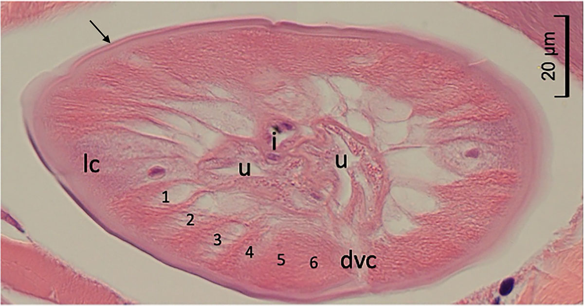
Histopathological examination. A cross section, cut slightly obliquely, of the worm found in the dermis of the patient, showing smooth cuticle (arrow), six somatic muscle cells (1–6) in a quadrant, lateral cord (lc) without internal cuticular projection, dorsal or ventral cord (dvc), and sections of intestine (i) and two uterine tubes (u) in the pseudocoelom.
Current usage metrics show cumulative count of Article Views (full-text article views including HTML views, PDF and ePub downloads, according to the available data) and Abstracts Views on Vision4Press platform.
Data correspond to usage on the plateform after 2015. The current usage metrics is available 48-96 hours after online publication and is updated daily on week days.
Initial download of the metrics may take a while.