Figure 3
Download original image
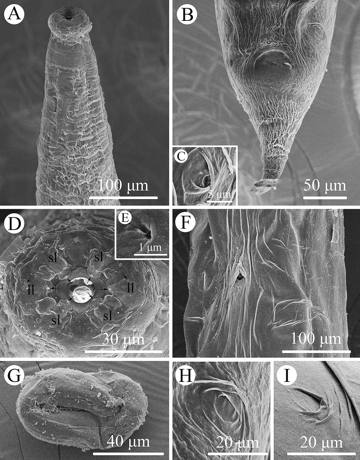
Scanning electron micrographs of Rhabdias macrocephalum n. sp. from Diploderma splendidum in China. A: anterior part of body, lateral view; B: tail, ventral view; C: magnified image of lateral cuticular pore; D: cephalic extremity (single papilla on each lip arrowed), apical view; E: magnified image of amphid; F: mid-body at level of vulva, sublateral view; G: egg with developed larva; H: magnified image of lateral cuticular pores on the tail; I: magnified image of lateral cuticular pores on the middle of body. Abbreviations: sl, submedian lip; ll, lateral lip.
Current usage metrics show cumulative count of Article Views (full-text article views including HTML views, PDF and ePub downloads, according to the available data) and Abstracts Views on Vision4Press platform.
Data correspond to usage on the plateform after 2015. The current usage metrics is available 48-96 hours after online publication and is updated daily on week days.
Initial download of the metrics may take a while.