Figure 5
Download original image
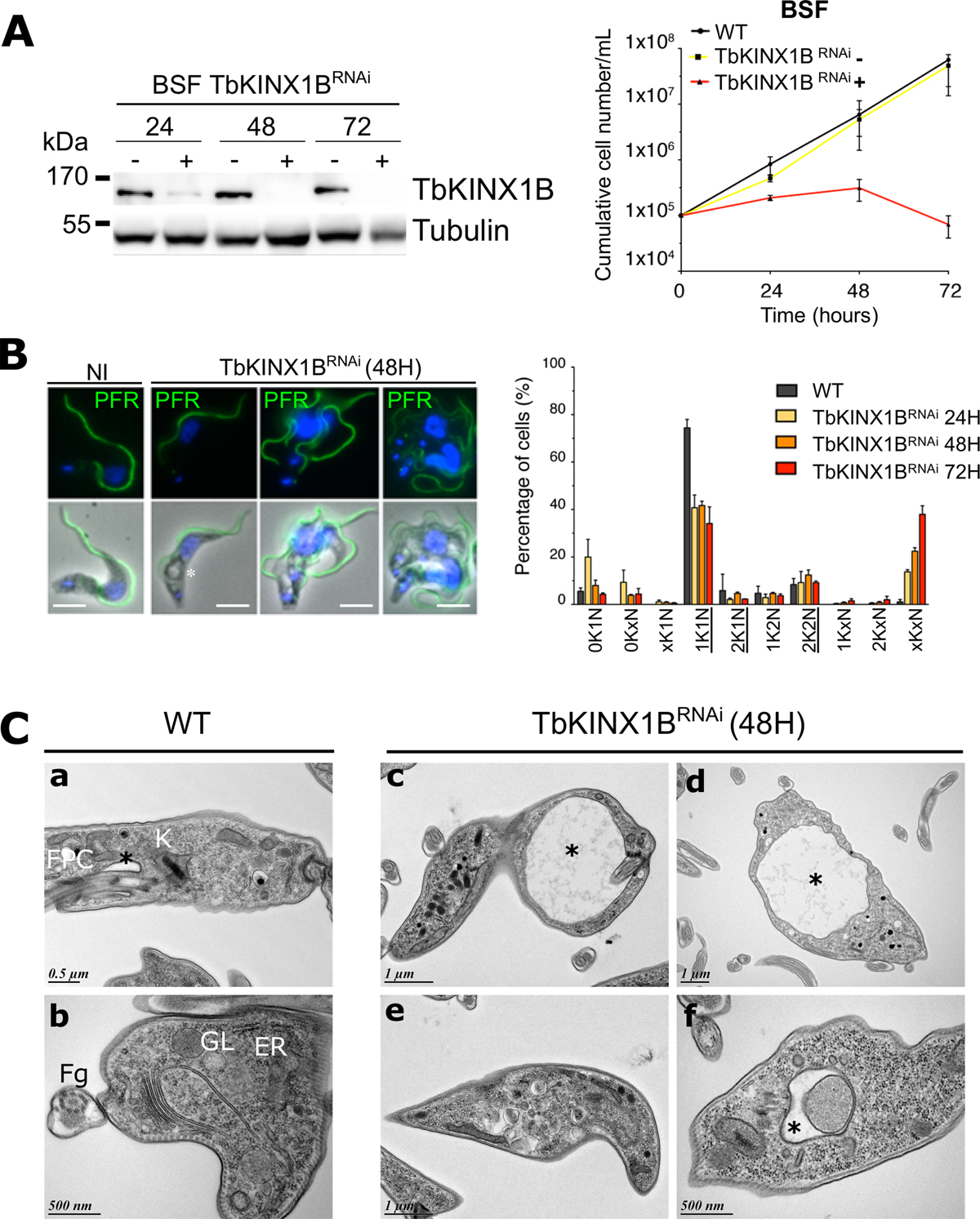
TbKINX1B is essential in bloodstream forms. A. Left panel, western blot of BSF whole cell protein extracts from TbKINX1BRNAi cell line in the absence (−) or presence (+) of tetracycline at 24 h, 48 h and 72 h using anti-TbKINX1B, and anti-tubulin (TAT1) as a loading control. Right panel, down-regulation of TbKINX1B in BSF (+) inhibits cell growth after 48 h of induction, as compared to non-induced cells (−) (n = 3). B. Left panel, IFA micrographs of WT and of 48 h induced TbKINX1BRNAi BSF cell lines. Immunofluorescence shows that down-regulation of TbKINX1B leads to multi-flagellated and multinucleated cells. Flagella were labelled with anti-PFR2 (green), and kinetoplasts and nuclei were DAPI-stained (blue). A large FP or vacuole is marked with an asterisk. Scale bars 5 μm. Right panel, quantification of cell division phenotypes in WT and in TbKINX1BRNAi cell lines after 24 h, 48 h and 72 h of induction. Normal phenotypes are underlined (200 cells, n = 3). C. Transmission Electron Microscopy (TEM) thin-sections of WT BSF and of TbKINX1BRNAi induced for 48 h. The structural organization of WT cells reveal well-defined FP (black asterisk), FPC, Golgi (G), endoplasmic reticulum (ER), recycling endosomes (RE), glycosomes (GL), Flagellum (Fg), internal vesicles and kinetoplast (K) (a, b). TbKINX1B knock-down cells possess enlarged FP (black asterisk, c, d), with the disturbed endo-membrane organization (i.e., FP harboring dense material or enlarged) (e, f).
Current usage metrics show cumulative count of Article Views (full-text article views including HTML views, PDF and ePub downloads, according to the available data) and Abstracts Views on Vision4Press platform.
Data correspond to usage on the plateform after 2015. The current usage metrics is available 48-96 hours after online publication and is updated daily on week days.
Initial download of the metrics may take a while.