Figure 4
Download original image
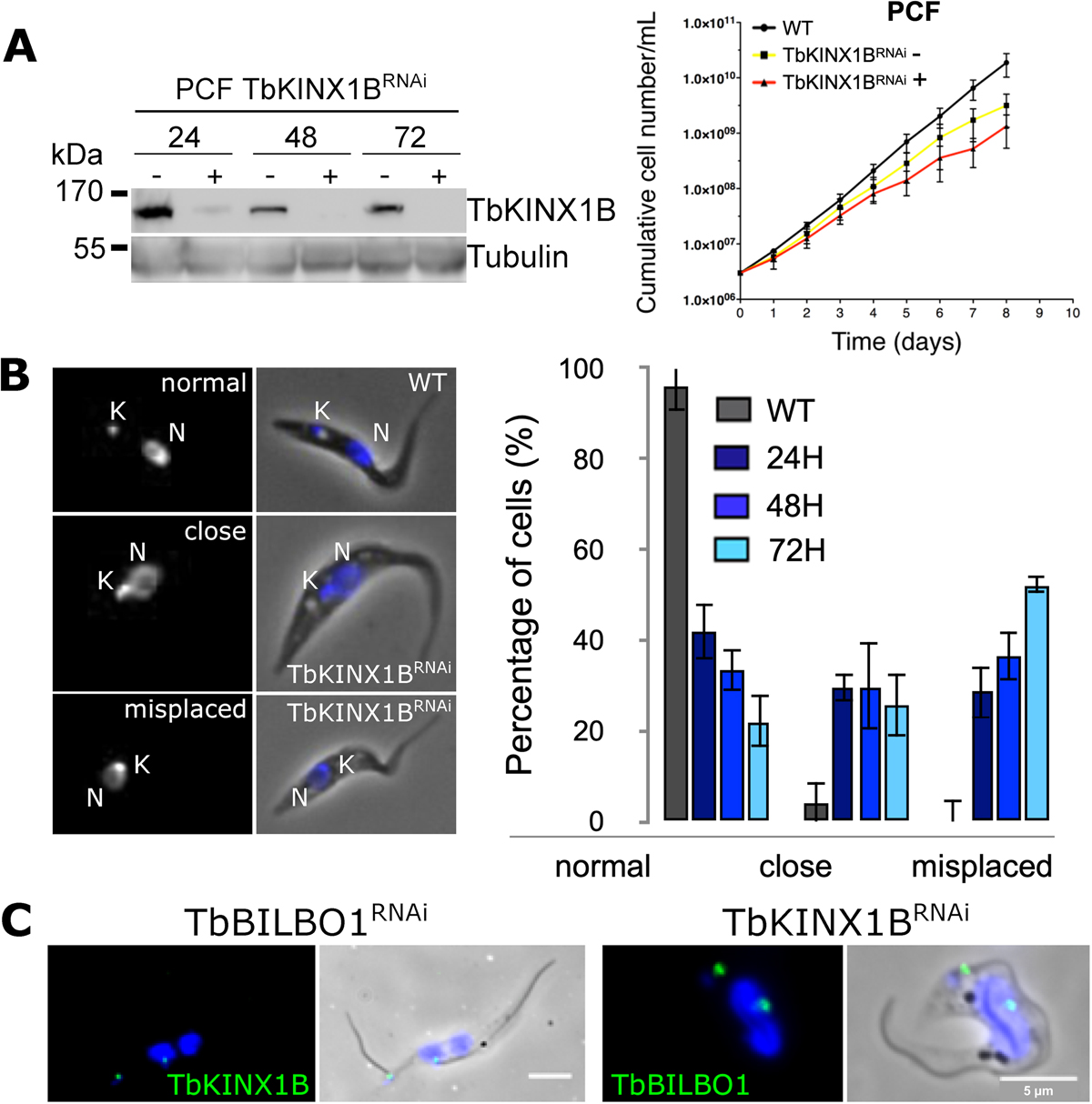
TbKINX1B is involved in basal body/FPC segregation in PCF. A. Left panel, Western blot analysis of whole cell protein extracts from PCF TbKINX1BRNAi cell line in the presence (+) or absence (−) of tetracycline after 24 h, 48 h or 72 h probed with anti-TbKINX1B, anti-TbBILBO1, and anti-Tubulin (TAT1) as a loading control (left panel). Right panel, PCF growth curves showing that induction of RNAi down-regulation of TbKINX1B (TbKINX1BRNAi) does not dramatically affect cell growth when compared to WT and non-induced cells (n = 3). B. Down-regulation of TbKINX1B induces misplaced kinetoplasts. Left panel, IFA micrographs from CSK preparation of WT (a) and TbKINX1BRNAi cell lines after 48 h of induction (b, c). Kinetoplasts (K) and nuclei (N) were stained with DAPI. Right panel, quantification of kinetoplast localization in 1K1N WT cells and in TbKINX1BRNAi cells after 24 h, 48 h and 72 h of induction. C. Left panel, immunolocalization of TbKINX1B (green) in TbBILBO1RNAi induced detergent-extracted cells. Right panel, immunolocalization of TbBILBO1 (green) in TbKINX1BRNAi induced detergent-extracted cells. Scale bars 5 μm.
Current usage metrics show cumulative count of Article Views (full-text article views including HTML views, PDF and ePub downloads, according to the available data) and Abstracts Views on Vision4Press platform.
Data correspond to usage on the plateform after 2015. The current usage metrics is available 48-96 hours after online publication and is updated daily on week days.
Initial download of the metrics may take a while.