Figure 3
Download original image
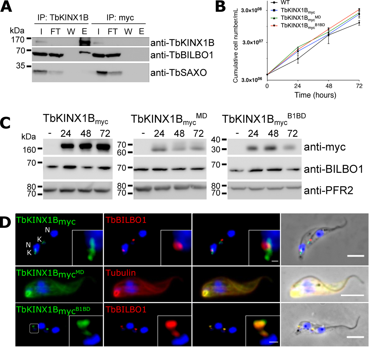
Microtubule and FPC localization of TbKINX1B depends on its motor domain and BILBO1-binding domain respectively. A. Immuno-precipitation of TbKINX1B and immuno-detection by western blotting of TbKINX1B and TbBILBO1 on input (I), flow-through (FT), wash (W), and elution (E) fractions. B. Induction of the expression of TbKINX1Bmyc, TbKINX1BmycMD or TbKINX1BmycB1BD does not affect PCF cell growth, as compared to WT (n = 3). C. Western blot of PCF WT cells and cells non-induced (–) or tetracycline-induced 24 h, 48 h, 72 h for the expression of TbKINX1Bmyc (151 KDa), TbKINX1BmycMD (62 KDa) or TbKINX1BmycB1BD (28 KDa). Expressed proteins were detected using anti-myc antibody and using PFR2 (L8C4) as a loading control. No alteration on TbBILBO1 protein levels was observed. D. Co-immunolabelling on detergent-extracted cells of TbKINX1Bmyc and TbKINX1BmycB1BD proteins (green) with TbBILBO1 (red), and of TbKINX1BmycMD (green) with Tubulin (red).
Current usage metrics show cumulative count of Article Views (full-text article views including HTML views, PDF and ePub downloads, according to the available data) and Abstracts Views on Vision4Press platform.
Data correspond to usage on the plateform after 2015. The current usage metrics is available 48-96 hours after online publication and is updated daily on week days.
Initial download of the metrics may take a while.