Figure 2
Download original image
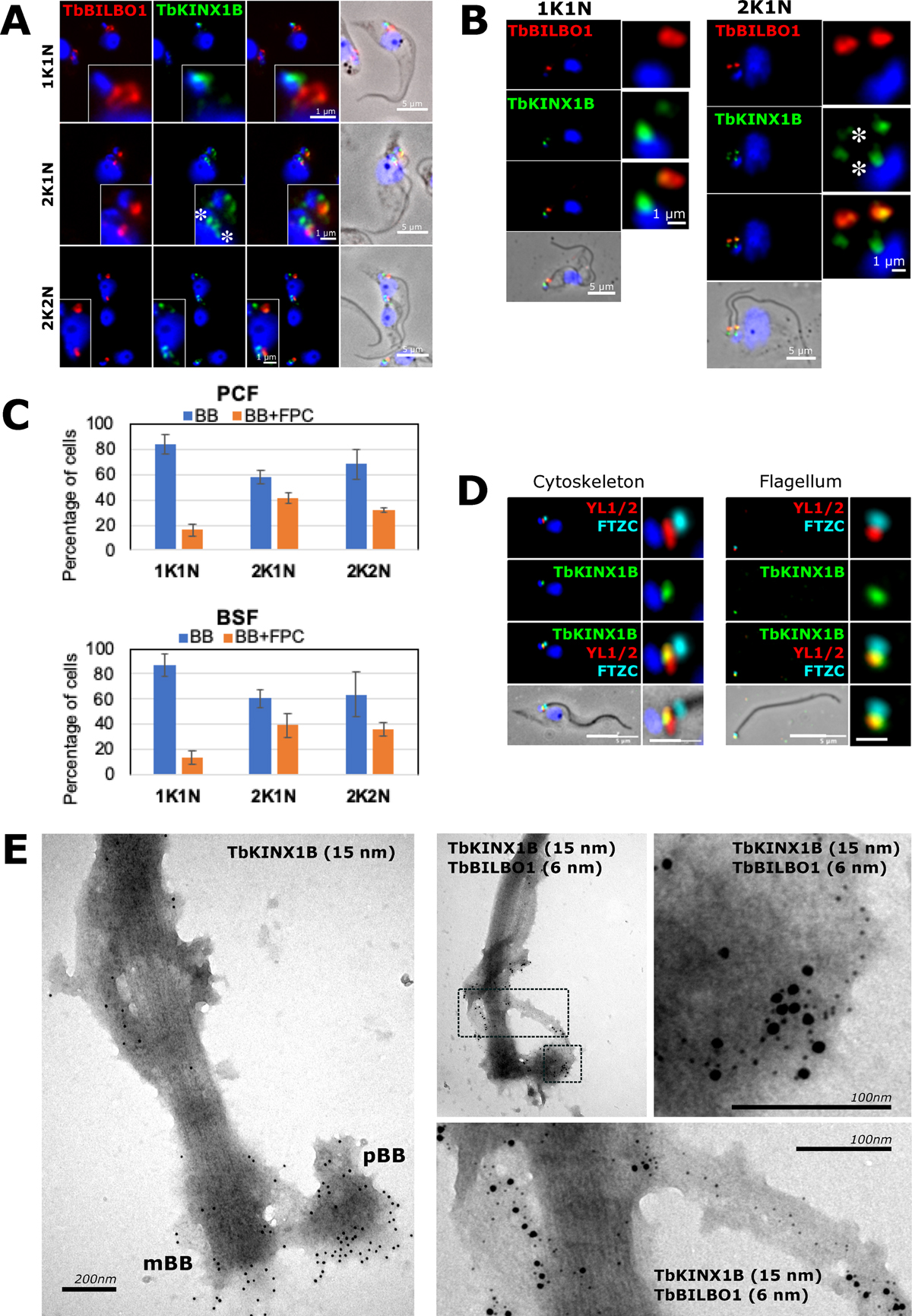
TbKINX1B displays a dual localization. A. Co-immunolabeling of TbKINX1B and TbBILBO1 on PCF detergent-extracted cells using anti-BILBO1 (red) and anti-TbKINX1B (green). B. Co-immunolabeling of TbKINX1B and BILBO1 on BSF detergent-extracted cells using anti-BILBO1 (red) and anti-TbKINX1B (green). C. Quantification of TbKINX1B BB and/BB+FPC localization in PCF and BSF from immunofluorescence experiments shown in A and B. BB localization (white bars) or BB and FPC localization (grey bars) were quantified at different cell cycle stages (200 cells, n = 3). D. Co-immunolabeling of TbKINX1B (green) and the basal bodies marker YL1/2 (red) and the transition zone marker FTZC (cyan) on detergent-extracted PCF cells cytoskeleton and flagellum. E. Immuno-TEM using PCF isolated flagella that were labelled with anti-TbKINX1B (15 nm gold particles), and co-immunolabeled with anti-TbKINX1B (15 nm gold) and anti-TbBILBO1 (6 nm gold). The images show positive labelling of the protein at the basal bodies (pBB, mBB), Flagellar Pocket Collar (FPC) and at the MTQ. The zoom image shows the TbBILBO1 6 nm gold beads on the MTQ and the colocalization of TbKINX1B and TbBILBO1. Scale bars in A, B and C represent 5 μm, and 1 μm in insets.
Current usage metrics show cumulative count of Article Views (full-text article views including HTML views, PDF and ePub downloads, according to the available data) and Abstracts Views on Vision4Press platform.
Data correspond to usage on the plateform after 2015. The current usage metrics is available 48-96 hours after online publication and is updated daily on week days.
Initial download of the metrics may take a while.