Figure 2
Download original image
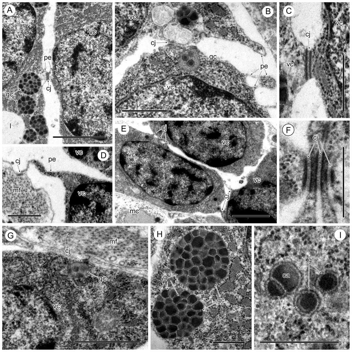
Intercellular junctions of the vitelline cells in Sanguinicola sp. 1 from pike. (A) Between two maturing vitelline cells, note the short cytoplasmic extension of both cells at the contact point. Scale bar = 2 μm. (B) Between two vitelline cells at an early stage of maturation, note the different kinds of pseudopodia-like extensions of the surface cytoplasm. Scale bar = 1 μm. (C, F) Detail of the junctions. Scale bars = 0.5 μm. (D) Between an immature vitelline cell and muscle fibres, note the long cytoplasmic extension of the vitelline cell at the contact point. Scale bar = 1 μm. (E) Between immature vitelline cells. Scale bar = 2 μm. (G) Between maturing vitelline cell and muscle fibres. Scale bar = 1 μm. (H) Vitelline clusters filled with vitelline globules. Scale bar = 1 μm. (I) Heterogeneous contents of the vitelline globules. Scale bar = 0.5 μm. Abbreviations: ca = central area of vitelline globule, cj = cell junction, g = vitelline globule, gc = globular cluster, mc = muscle cell, mf = muscle fibres, pe = pseudopodia-like extension, r = lucid rim of vitelline globule, vc = vitelline cell.
Current usage metrics show cumulative count of Article Views (full-text article views including HTML views, PDF and ePub downloads, according to the available data) and Abstracts Views on Vision4Press platform.
Data correspond to usage on the plateform after 2015. The current usage metrics is available 48-96 hours after online publication and is updated daily on week days.
Initial download of the metrics may take a while.