Figure 1
Download original image
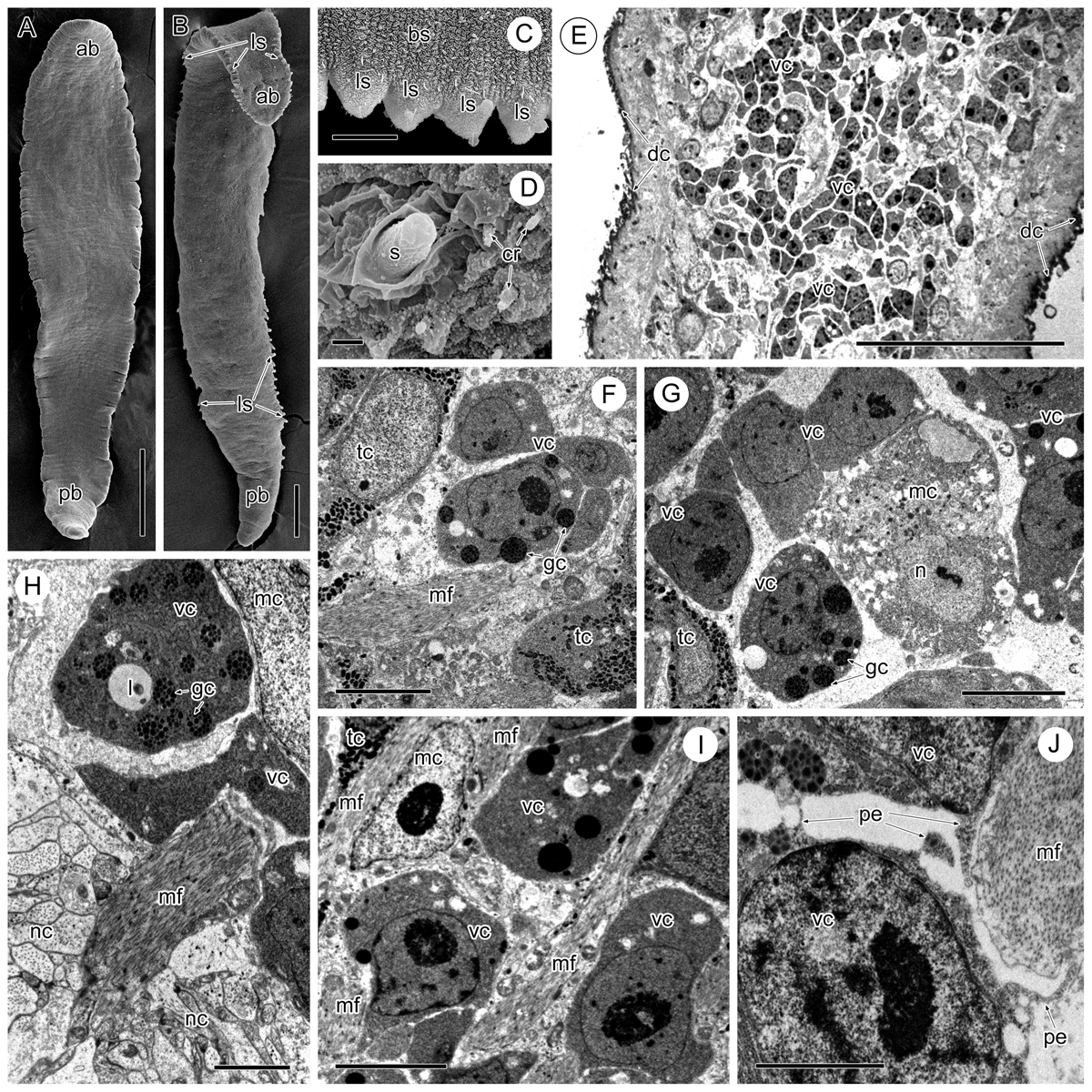
SEM images of Sanguinicola spp. (A–D) and TEM images of the vitelline cells of Sanguinicola sp. 1 from pike (E–J). (A) Sanguinicola sp. 1 from pike, note the absence of visible lateral spines along the body. Scale bar = 200 μm. (B) Sanguinicola sp. 2 from ides, note the presence of lateral spines along the body surface. Scale bar = 100 μm. (C) Lateral spines of Sanguinicola sp. 2. Scale bar = 10 μm. (D) Spine of Sanguinicola sp. 1 localized laterally in the anterior third of the body. Scale bar = 1 μm. (E) Longitudinal section through part of the body, note vitelline cells at different stages of maturation mixed with other cell types. Scale bar = 50 μm. (F) Vitelline cells surrounded by tegumentary cells and muscle fibres. Scale bar = 5 μm. (G) Vitelline cells mixed with muscle and tegumental cells. Scale bar = 5 μm. (H) Vitelline cells surrounded by a muscle cell, note the neighbouring nerve cord and muscle fibres. Scale bar = 2 μm. (I) Mixed vitelline cells, muscle cells and muscle fibres. Scale bar = 5 μm. (J) Pseudopodia-like extensions of a vitelline cell directed towards a neighbouring vitelline cell and muscle fibres. Scale bar = 2 μm. Abbreviations: ab = anterior part of body, bs = body surface, cr = ciliary receptor, dc = distal cytoplasm of tegument, gc = globular cluster, l = lipid droplet, ls = lateral spine, mc = muscle cell, mf = muscle fibres, n = nucleus, nc = nerve cord, pb = posterior part of body, pe = pseudopodium-like extension, s = spine, tc = tegumentary cell, vc = vitelline cell.
Current usage metrics show cumulative count of Article Views (full-text article views including HTML views, PDF and ePub downloads, according to the available data) and Abstracts Views on Vision4Press platform.
Data correspond to usage on the plateform after 2015. The current usage metrics is available 48-96 hours after online publication and is updated daily on week days.
Initial download of the metrics may take a while.