Figure 6
Download original image
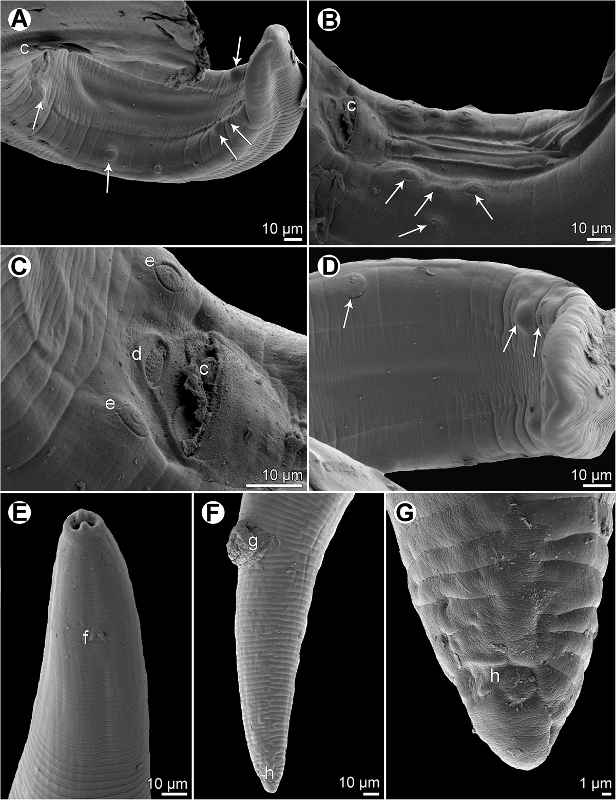
Ascarophis (Ascarophis) nasonis Machida, 1981 from Naso spp., scanning electron micrographs. (A) Tail of male, sublateral view (arrows indicate postanal papillae); (B) precloacal region of male, ventrolateral view (arrows indicate preanal papillae); (C) region of cloaca, subventral view; (D) posterior part of male tail, ventral view (arrows indicate postanal papillae); (E) anterior end of body, lateral view; (F) tail of female, lateral view; (G) posterior end of female tail, lateral view. (c) Cloacal aperture; (d) median depression on posterior cloacal lip; (e) papillae of first postanal pair; (f) deirid; (g) anus; (h) phasmid.
Current usage metrics show cumulative count of Article Views (full-text article views including HTML views, PDF and ePub downloads, according to the available data) and Abstracts Views on Vision4Press platform.
Data correspond to usage on the plateform after 2015. The current usage metrics is available 48-96 hours after online publication and is updated daily on week days.
Initial download of the metrics may take a while.