Figure 1.
Download original image
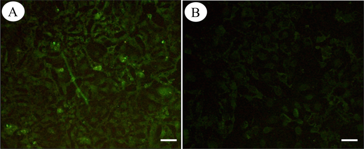
Immunofluorescence staining of HSP40 in HEK293 cells. HEK293 cells were transfected with pVAX1-HSP40 or pVAX1. At 72 h after transfection, the cells were fixed, permeabilized, and incubated with anti-T. gondii primary antibody. The cells were then incubated with fluorescein-conjugated secondary antibody and observed under a fluorescence microscope. The experiment was repeated three times, yielding similar results. The cells transfected with pVAX1-HSP40 showed the immunofluorescence of HSP40 proteins in the cytoplasm (A), whereas the cells transfected with the empty pVAX1 plasmid displayed little cellular immunofluorescence (B). Scale bar: 10 μm.
Current usage metrics show cumulative count of Article Views (full-text article views including HTML views, PDF and ePub downloads, according to the available data) and Abstracts Views on Vision4Press platform.
Data correspond to usage on the plateform after 2015. The current usage metrics is available 48-96 hours after online publication and is updated daily on week days.
Initial download of the metrics may take a while.