Figure 3
Download original image
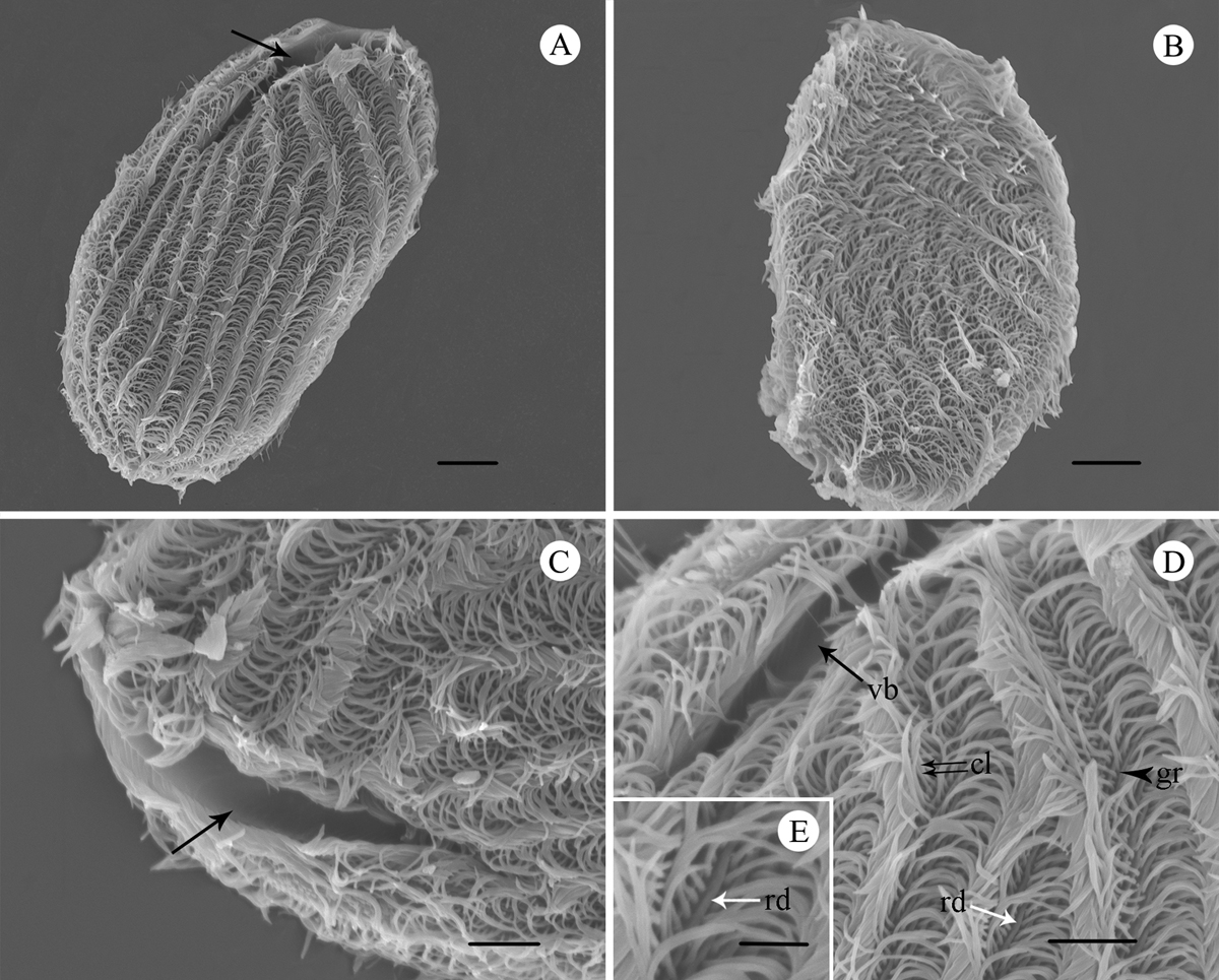
SEM images of B. grimi. A. Overview of the ventral-left side (oral side), showing the general form, vestibulum (arrow) and uniformly arranged cilia. Scale bar = 10 µm. B. Overview of the right side, showing the body surface is partially flattened and thickly ciliated. Scale bar = 10 µm. C. Ventral-left view of the “V”-shaped vestibulum (arrow). Scale bar = 5 µm. D. The left anterior area of ciliate, showing the vestibulum (vb), an interkinetal ridge (rd), the groove (gr) and the cilia (cl) extending from grooves and are close to one another. Scale bar = 5 µm. E. Selected enlargement of Figure 3D, showing a ridge (rd) between cilia. Scale bar = 2 µm.
Current usage metrics show cumulative count of Article Views (full-text article views including HTML views, PDF and ePub downloads, according to the available data) and Abstracts Views on Vision4Press platform.
Data correspond to usage on the plateform after 2015. The current usage metrics is available 48-96 hours after online publication and is updated daily on week days.
Initial download of the metrics may take a while.