Figure 2.

Download original image
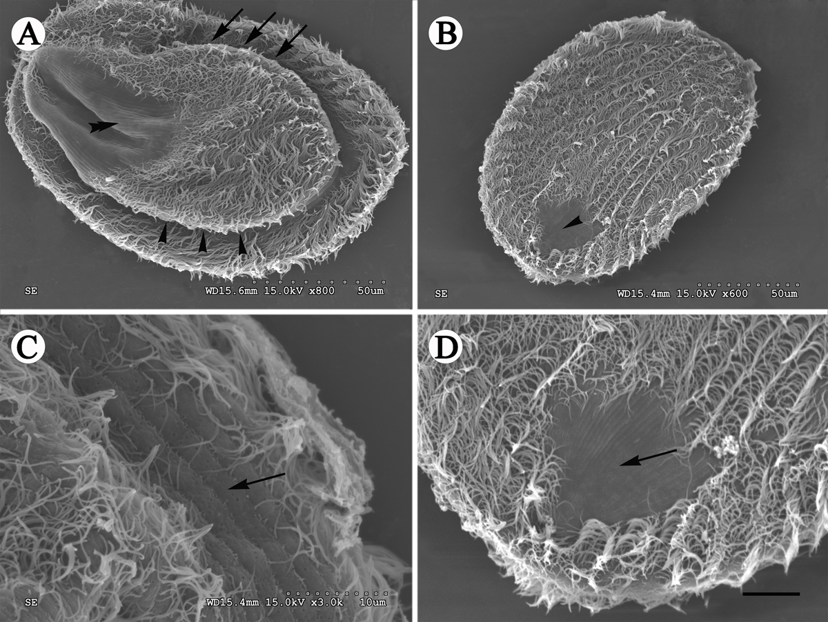
SEM images of Sicuophora multigranularis. (A) Left side view showing the oral groove (arrows) and its convex left side as well as the naked region without cilia at the posterior body region (double arrowheads), and two very different sides. Scale bar = 50 μm. (B) Right side view, showing the densely arranged ciliary rows and the glabrous region without cilia in the posterior body region. Scale bar = 50 μm. (C) Detail of specimen shown in Fig. 2A. Note the sparely arranged cilia on the cell margin. Scale bar = 10 μm. (D) Detail of specimen shown in Fig. 2B. There is a conspicuous naked area on the right side near the posterior body end. Scale bar = 10 μm.
Current usage metrics show cumulative count of Article Views (full-text article views including HTML views, PDF and ePub downloads, according to the available data) and Abstracts Views on Vision4Press platform.
Data correspond to usage on the plateform after 2015. The current usage metrics is available 48-96 hours after online publication and is updated daily on week days.
Initial download of the metrics may take a while.


