Figure 3.
Download original image
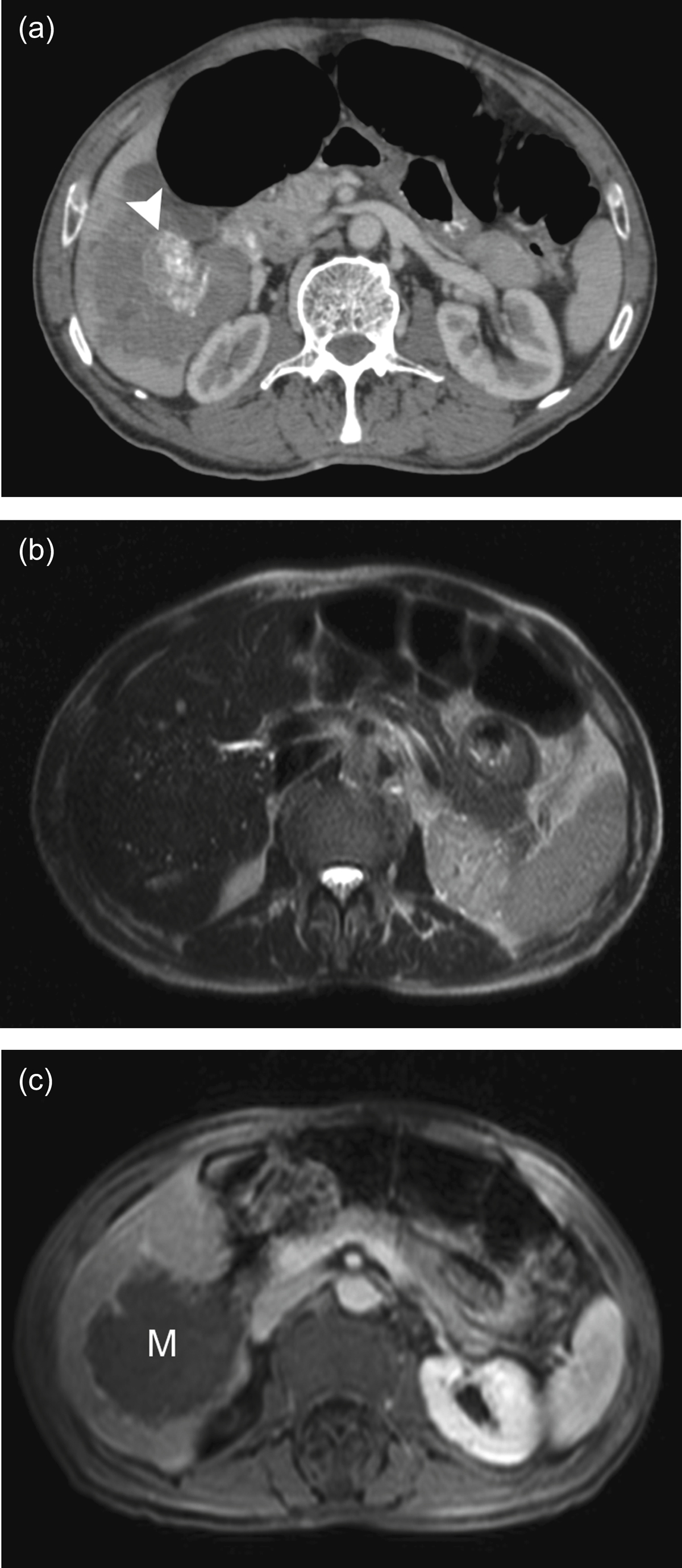
A 78-year-old male patient previously operated for gastric cancer was found to have a mass in the right lobe of the liver. (a) Axial contrast-enhanced CT examination shows an irregularly bordered, hypovascular, heterogeneous mass lesion in the right lobe. Focal calcific areas (arrowhead) in the center of the lesion can be clearly discerned. (b) On an axial T2W MRI image, the mass is not clearly discernible from the normal liver parenchyma. However, many hyperintense, small cysts can be seen at the peripheral zones of the lesion. (c) A postcontrast axial MRI image demonstrates that the mass (M) did not show prominent contrast uptake with the exception of a weak, peripheral contrast uptake. With an elevated CA 19-9 level, the patient underwent core biopsy to exclude a malignancy, and the result indicated the diagnosis of AE.
Current usage metrics show cumulative count of Article Views (full-text article views including HTML views, PDF and ePub downloads, according to the available data) and Abstracts Views on Vision4Press platform.
Data correspond to usage on the plateform after 2015. The current usage metrics is available 48-96 hours after online publication and is updated daily on week days.
Initial download of the metrics may take a while.