Figure 2.
Download original image
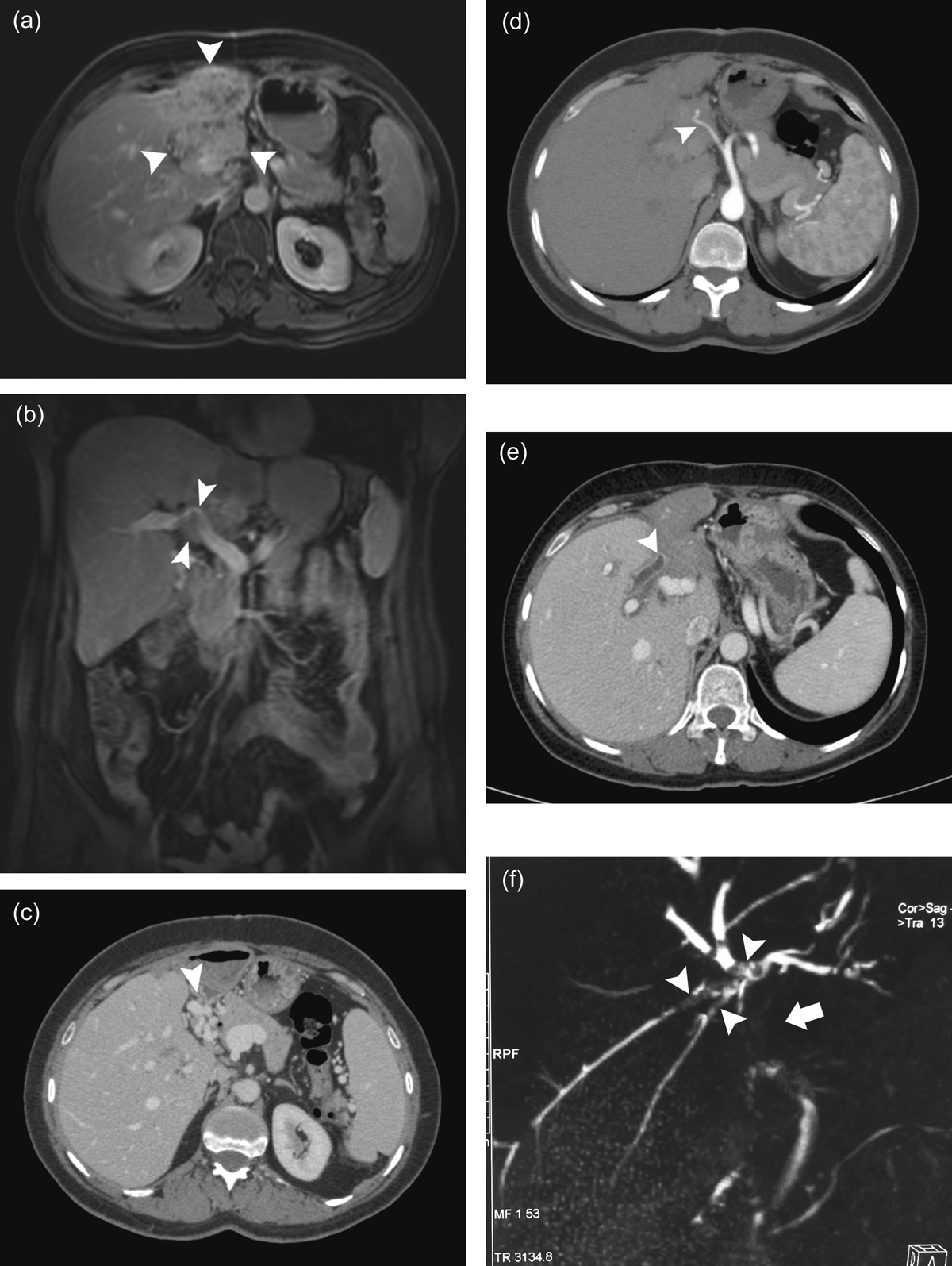
A 51-year-old female patient presented with back pain. (a) Abdominal MRI with contrast demonstrates a mass lesion (arrowheads) with indistinct borders, which completely fills the left lobe of the liver with concurrent atrophy and shows diffuse heterogeneous enhancement. (b) Postcontrast coronal MRI image demonstrates portal vein invasion and narrowing (arrowheads). (c) Many collateral veins (arrowhead) that developed at the hilus secondary to portal vein invasion are demonstrated by portal phase CT examination. (d) An arterial phase axial CT image reveals the hepatic artery wrapped by the mass (arrowhead). (e) Contrast-enhanced abdominal CT examination shows that the contrast-enhanced, thickened common bile duct is interrupted within the lesion (arrowhead). (f) Filling defects due to the bile ducts invaded by the mass (arrowheads) and the interruption of the common bile duct (arrow) are more clearly visualized by MRCP images. Cholangiocarcinoma was considered in the differential diagnosis; however, the core biopsy result was consistent with AE.
Current usage metrics show cumulative count of Article Views (full-text article views including HTML views, PDF and ePub downloads, according to the available data) and Abstracts Views on Vision4Press platform.
Data correspond to usage on the plateform after 2015. The current usage metrics is available 48-96 hours after online publication and is updated daily on week days.
Initial download of the metrics may take a while.