Figure 5
Download original image
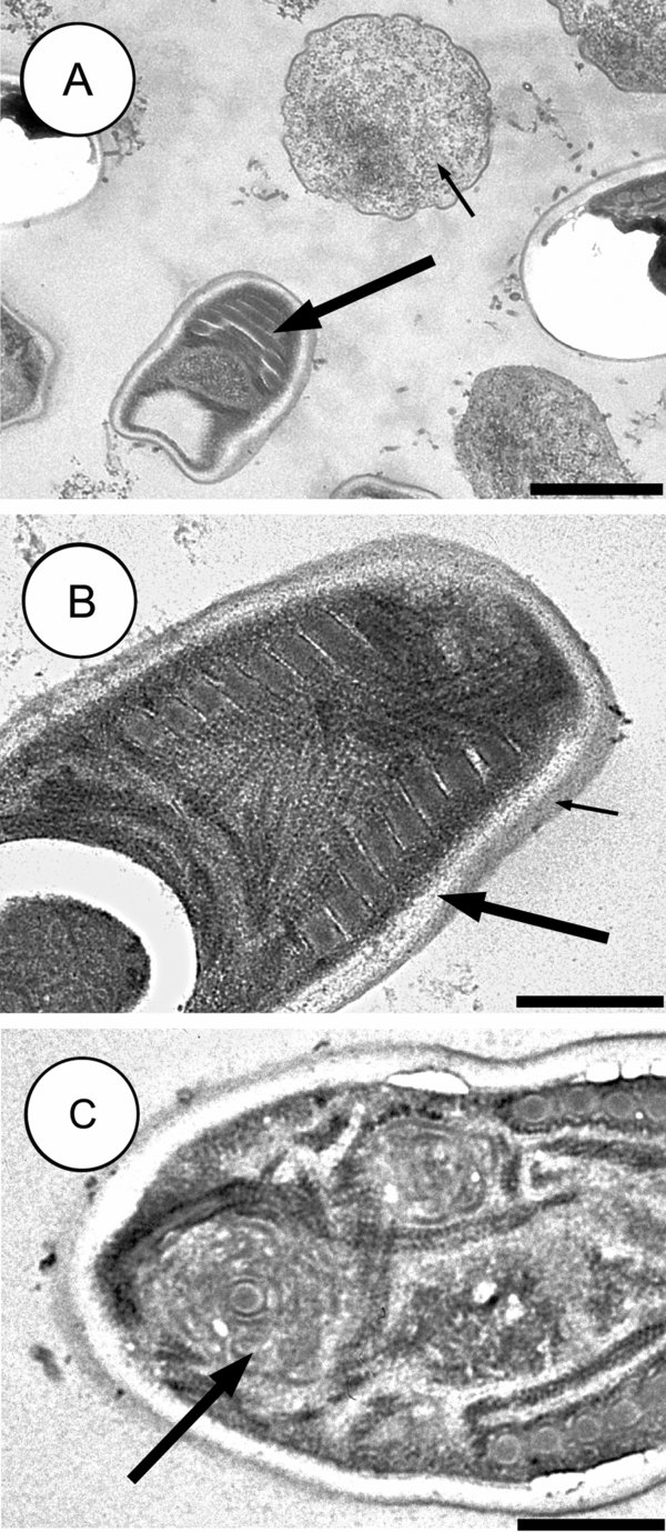
Dictyocoela diporeiae n. sp., transmission electron micrograph of the microsporidian infecting Diporeia sp. in Lake Superior. Notice (A) the meront (small arrow) and mature spore (large arrow), (B) spore wall composed of a thick electron-lucent endospore (large arrow) overlaid with a thinner electron-dense exospore (small arrow), and (C) lamellar polaroplast composed of ordered concentric membranes surrounding the polar filament (large arrow). Scale bars: A = 1000 nm, B–C = 500 nm.
Current usage metrics show cumulative count of Article Views (full-text article views including HTML views, PDF and ePub downloads, according to the available data) and Abstracts Views on Vision4Press platform.
Data correspond to usage on the plateform after 2015. The current usage metrics is available 48-96 hours after online publication and is updated daily on week days.
Initial download of the metrics may take a while.