Figure 3.
Download original image
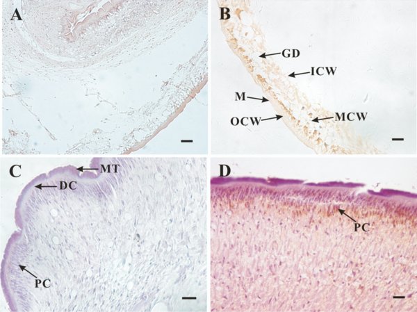
Immunolocalisation of TpFABP in T. pisiformis tapeworm and cysticercus. The yellowish-brown tint shows the TpFABP protein location. (A) negative sera in cysticercus; (B) antisera in cysticercus; (C) negative sera in adult tapeworm; (D) antisera in adult tapeworm. Arrows indicate the areas of the parasite: MT, microthrix; DC, distal cytoplasm; PC, perinuclear cytoplasm; GD, gathering duct; M, microtrichia; ICW, inside the layer of cystic wall; OCW, outer layer of cystic wall; MCW, middle layer of cystic wall. Scale bars: 20 μm.
Current usage metrics show cumulative count of Article Views (full-text article views including HTML views, PDF and ePub downloads, according to the available data) and Abstracts Views on Vision4Press platform.
Data correspond to usage on the plateform after 2015. The current usage metrics is available 48-96 hours after online publication and is updated daily on week days.
Initial download of the metrics may take a while.