Fig. 1.
Download original image
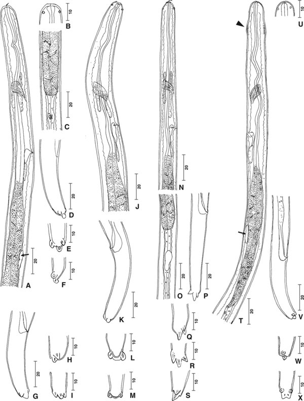
Infective larvae of Cercopithifilaria species from ticks collected from the Japanese serow. (A–F) Type A larva. A. Anterior part, right lateral view. Female genital primordium, arrow. B. Head. C. Oesophageal-intestinal junction. D. Tail, right lateral view at anus. E. Caudal end, ventral view. F. Caudal end, left lateral view. (G–I) Type B larva. G. Tail, right lateral view. H. Caudal end, lateral view. I. Caudal end, ventral view. (J–M) Type C larva. J. Anterior part, right lateral view. K. Tail, left lateral view. L-M. Caudal end, ventral view. (N–S) Type D larva. N. Anterior part, right lateral view. O. Oesophageal-intestinal junction. *Male genital primordium. P. Tail, right lateral view at anus; ventral view at the end. Q. Caudal end, right lateral view. R. Caudal end, ventral view. S. Caudal end, left lateral view. (T–X) Type E larva. T. Anterior part, left lateral view. Cervical swelling, arrowhead; female genital primordium, arrow. U. Head. V. Tail, right lateral view. W. Caudal end, lateral view. X. Caudal end, ventral view. Scale bars: micrometres.
Current usage metrics show cumulative count of Article Views (full-text article views including HTML views, PDF and ePub downloads, according to the available data) and Abstracts Views on Vision4Press platform.
Data correspond to usage on the plateform after 2015. The current usage metrics is available 48-96 hours after online publication and is updated daily on week days.
Initial download of the metrics may take a while.