Figs 1–16.
Download original image
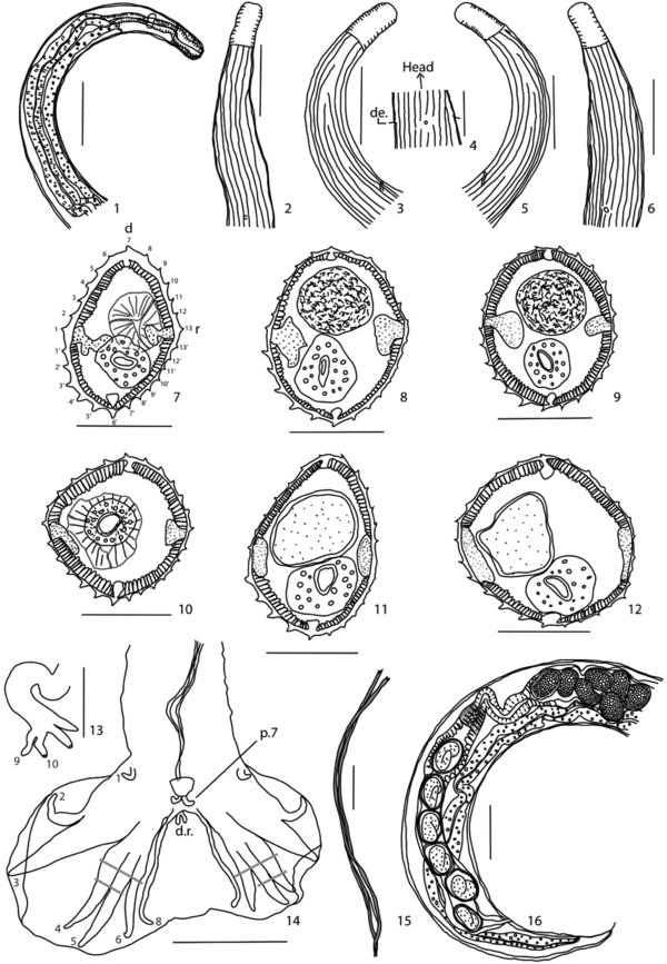
Heligmosomoides neopolygyrus Asakawa and Ohbayashi, 1986, in Apodemus peninsulae, from China: 1-6, male, anterior extremity, 1, right lateral view, 2-3, origin of cuticular ridges, 2, dorsal view, 3, right lateral view, 4, detail of excretory pore and deirids, ventral view, 5-6, origin of cuticular ridges, 5, left lateral view, 6, sub-ventral view; 7-12, transverse sections of body, 7-9, male, 7, at level of esophago-intestinal junction, 8, at mid-body, 9, within distal fifth, 10-12, female, 10, at level of esophago-intestinal junction, 11, at midbody, 12, within distal fifth; 13-15, male, 13, dorsal ray with rays 9 and 10, ventral view, 14, caudal bursa, ventral view, 15, spicules, in situ, ventral view; 16, female, posterior extremity, right lateral view.
Scale bar: Figs 1-3, 5-6, 14, 16: 100 μm. Figs 4, 7-12, 15: 50 μm. Fig. 13: 20 μm. Abbreviations: de: deirids, r: right side, d: dorsal side, d.r.: dorsal ray, p.7: papillae 7. Transverse sections are oriented and numbered as in Fig. 7.
Current usage metrics show cumulative count of Article Views (full-text article views including HTML views, PDF and ePub downloads, according to the available data) and Abstracts Views on Vision4Press platform.
Data correspond to usage on the plateform after 2015. The current usage metrics is available 48-96 hours after online publication and is updated daily on week days.
Initial download of the metrics may take a while.