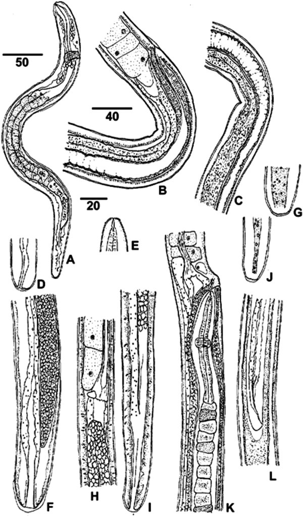
Fig. 2.
Download original image
Morphology of developing Trichosomoides nasalis in Arvicanthis niloticus.
A. First-stage larva, three days old, showing nerve ring, muscular oesophagus, long stichosome and short intestine, left lateral view. B-D. Female larva 2,800 μm long at very beginning of fourth moult; B. Oesophageal-intestinal junction and vulva (under exuvial sheath), vagina and distal part of uterus, right lateral view; C. Continuation of B, with uterus and intestine; D. Posterior extremity with rectum, lateral view. E-G. Female larva, 2,350 μm long; E. Head and stylet; F. Posterior region with ovary, intestine and rectum, and slight terminal exuvial sheath, dorso-ventral view; G. Bacillary band at posterior extremity, lateral view. H. Adult male 1,750 μm long, oesophageal-intestinal junction 1,050 μm from head. I. Same male, posterior extremity with intestine (on left), ejaculatory duct and cloaca, lateral view. J. Bacillary band at posterior extremity of a male, lateral view. K-L. Adult female 5,000 μm long with male; K. Male anterior part, its head oriented to vulva (note the stichocytes dark or clear); L. Male posterior part in uterus.
A-G: worms from abdominal wall, H-L: worms from nasal mucosa. All worms recovered from series of re-infected rodents, except the first-stage larva.
Scale bars in μm: A, K, L, 50; B, C, 40; D-J, 20.
Current usage metrics show cumulative count of Article Views (full-text article views including HTML views, PDF and ePub downloads, according to the available data) and Abstracts Views on Vision4Press platform.
Data correspond to usage on the plateform after 2015. The current usage metrics is available 48-96 hours after online publication and is updated daily on week days.
Initial download of the metrics may take a while.