


Figure 6
Download original image
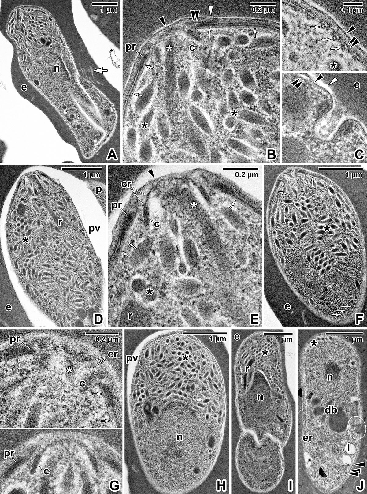
Subcellular organisation of Dactylosoma cf. ranarum SK gamonts. A) Anterior region of an immature gamont. B) Apical end of the gamont shown in A. C) Pellicle details showing subpellicular microtubules (top micrograph) and an active micropore with membrane structures filling its invaginated part (bottom micrograph). D) Anterior region of a more advanced stage of an immature gamont. E) Detail of the apical end of the gamont shown in D. F) Anterior region of a maturing gamont. G) Detailed views of the apical ends of maturing gamonts (top micrograph corresponds to the gamont shown in F). H) Anterior region of a mature gamont with a prominent nucleus and numerous micronemes. I) Longitudinal section of a recurved mature gamont in its folding region. J) Mid-posterior part of a mature gamont with a micropore (encircled) and several dense bodies. Transmission electron microscopy. Black arrowhead – parasite plasma membrane, black asterisk – microneme(s), c – conoid, cr – preconoidal rings, db – dense bodies, double black arrowhead – inner membrane complex, e – erythrocyte, er – rough endoplasmic reticulum, n – nucleus, pr – polar ring(s), pv – inner space of parasitophorous vacuole, r – rhoptry, small white arrow – subpellicular microtubule, white arrow – extensions of the parasitophorous vacuole membrane into the erythrocyte cytoplasm, white arrowhead – parasitophorous vacuole, white asterisk – rhoptry duct.
Current usage metrics show cumulative count of Article Views (full-text article views including HTML views, PDF and ePub downloads, according to the available data) and Abstracts Views on Vision4Press platform.
Data correspond to usage on the plateform after 2015. The current usage metrics is available 48-96 hours after online publication and is updated daily on week days.
Initial download of the metrics may take a while.