


Figure 5
Download original image
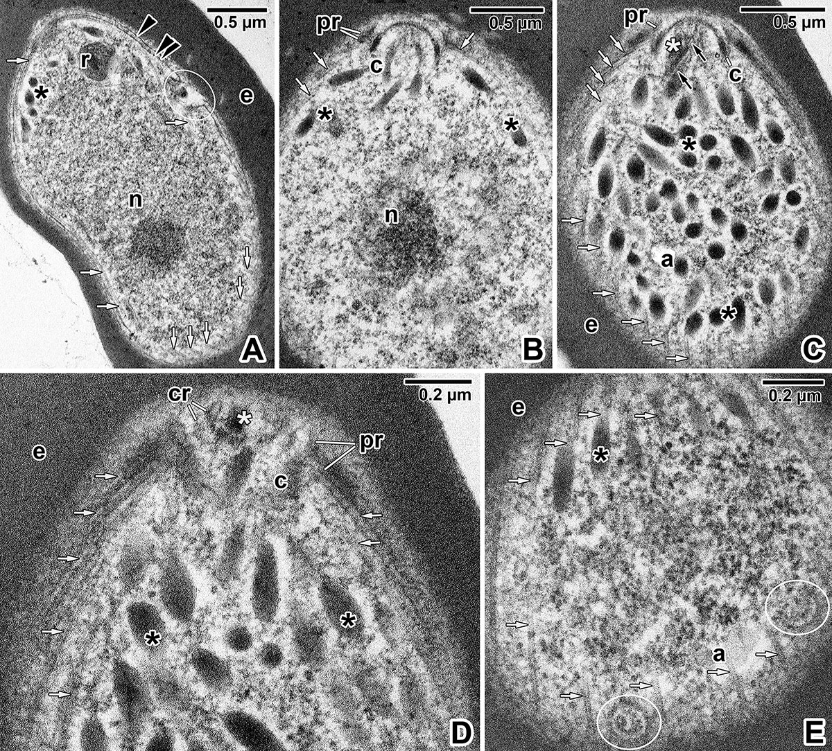
Subcellular organisation of Dactylosoma cf. ranarum SK secondary merozoites. A) Tangential section through a merozoite following erythrocyte invasion. Note the micropore (encircled) in its anterior region. B–C) Apical end of a secondary merozoite in mid-longitudinal (B) and superficial (C) sections. D–E) Tangentially sectioned anterior end of secondary merozoite showing the apical complex (D) and subpellicular microtubules with circular structures (encircled in E). Transmission electron microscopy. a – amylopectin granule, black arrowhead – parasite plasma membrane, black asterisk – microneme(s), c – conoid, cr – preconoidal rings, double black arrowhead – inner membrane complex, e – erythrocyte, n – nucleus, pr – polar ring(s), r – rhoptry, small black arrow – intraconoidal microtubule, small white arrow – subpellicular microtubule, white asterisk – rhoptry duct.
Current usage metrics show cumulative count of Article Views (full-text article views including HTML views, PDF and ePub downloads, according to the available data) and Abstracts Views on Vision4Press platform.
Data correspond to usage on the plateform after 2015. The current usage metrics is available 48-96 hours after online publication and is updated daily on week days.
Initial download of the metrics may take a while.