Figure 1
Download original image
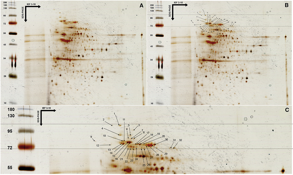
2-DE gel obtained with T. gondii lysate antigen after silver nitrate staining. A. All proteins from T. gondii RH strain tachyzoites were separated by isoelectric focusing (pH 3–10), followed by SDS-PAGE electrophoresis on polyacrylamide gel (12% acrylamide), and then revealed by silver nitrate staining. B. Delineation of the IgM triplet zone between 75 and 100 kDa and numbering of all protein spots present. C. Close-up of the protein spots in the IgM triplet zone.
Current usage metrics show cumulative count of Article Views (full-text article views including HTML views, PDF and ePub downloads, according to the available data) and Abstracts Views on Vision4Press platform.
Data correspond to usage on the plateform after 2015. The current usage metrics is available 48-96 hours after online publication and is updated daily on week days.
Initial download of the metrics may take a while.