Figure 3
Download original image
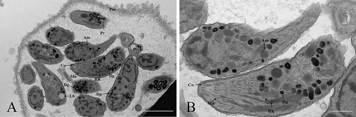
Morphology of tachyzoites of TgFurSealCHn1 in cell culture under transmission electron microscopy. A: A group of TgFurSealCHn1 tachyzoites within the parasitophorous vacuolar membrane. B: Magnified image of A. Am: amylopectin granule, Co: conoid, Dg: electron-dense granule, Lb: lipid, Mn: microneme, Nu: nuclei of daughter tachyzoites, Pm: parasitophorous vacuolar membrane, Pv: parasitophorous vacuole, Rh: rhoptry. Bar = 2 μm.
Current usage metrics show cumulative count of Article Views (full-text article views including HTML views, PDF and ePub downloads, according to the available data) and Abstracts Views on Vision4Press platform.
Data correspond to usage on the plateform after 2015. The current usage metrics is available 48-96 hours after online publication and is updated daily on week days.
Initial download of the metrics may take a while.