Figure 2
Download original image
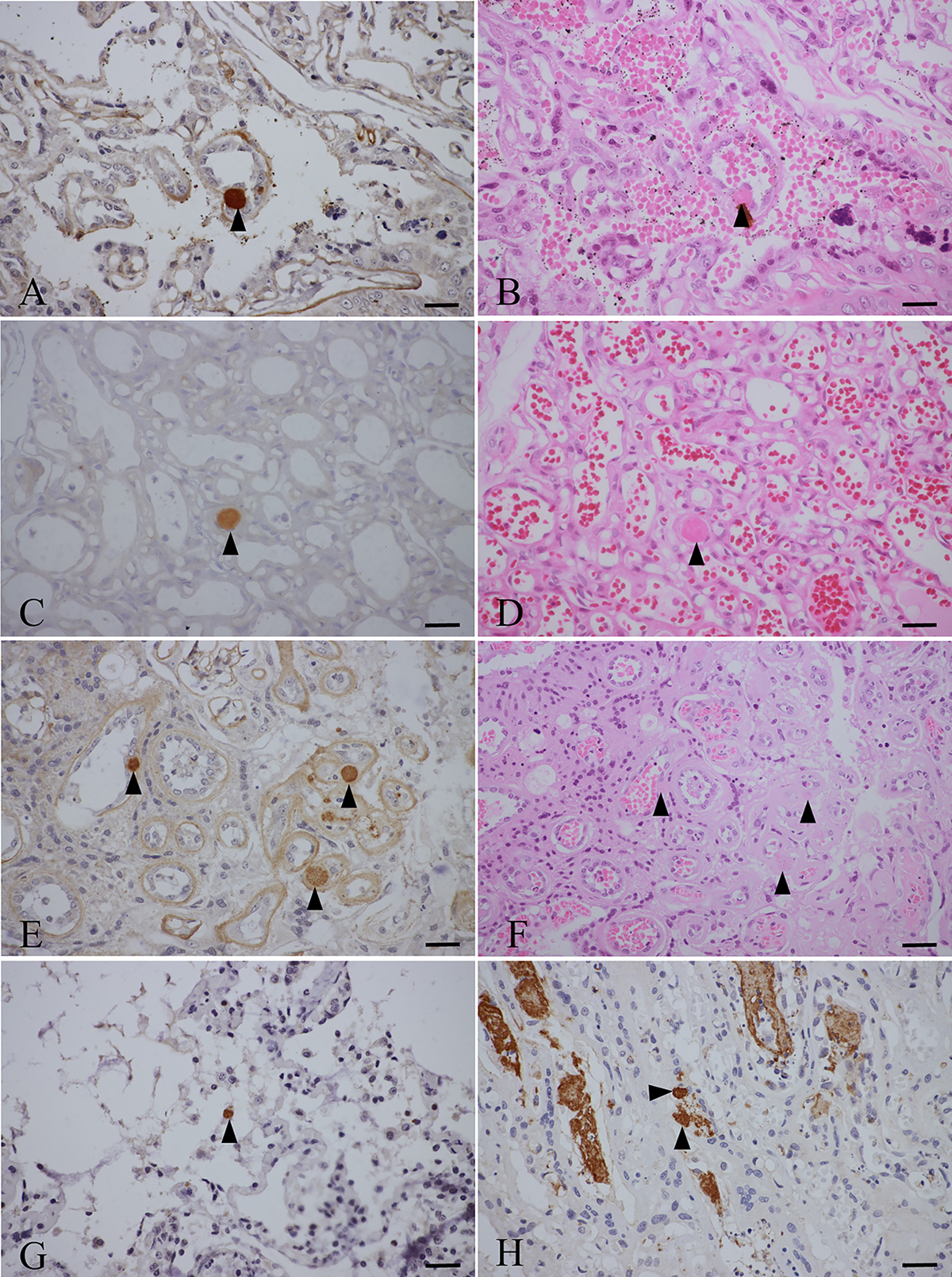
Morphology of T. gondii in the tissues of the fur seals. A–D: T. gondii antibody positive reaction (trigon) in the placentas of fur seal #2. A and C show IHC staining, while B (P#3445) and D (P#3559) show HE staining of serial sections A and C, respectively. E, F: T. gondii antibody positive reaction (trigon) in placentas of fur seal #4. E shows IHC staining, and F (P#3568) shows HE staining of the E serial section. G: T. gondii antibody positive reaction in the lung of fur seal #7 (P#3814) shown by IHC staining (trigon). H: T. gondii antibody positive reaction in the placenta of fur seal #8 (P#3841) shown by IHC staining (trigon). Bar = 50 μm.
Current usage metrics show cumulative count of Article Views (full-text article views including HTML views, PDF and ePub downloads, according to the available data) and Abstracts Views on Vision4Press platform.
Data correspond to usage on the plateform after 2015. The current usage metrics is available 48-96 hours after online publication and is updated daily on week days.
Initial download of the metrics may take a while.