Figure 2
Download original image
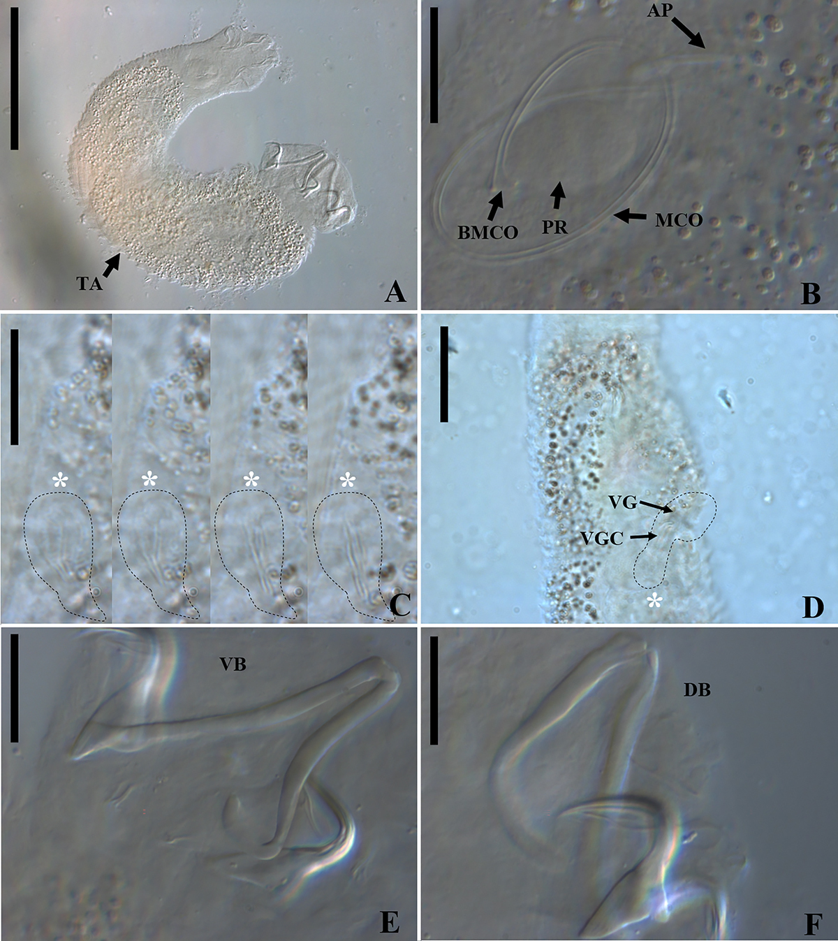
Light photomicrographs of Demidospermus paranaensis. A – whole worm in ventral view (CHIOC37255b) showing the tegument annulations (TA). B – MCO: accessory piece (AP); base of the MCO (BMCO); prostatic reservoir (PR) (CHIOC37255b). C – sclerotized distal portion of the deferens duct, observed at varying depths, from left to right, ventral to dorsal: asterisk indicates the same topographical region in D. D – vagina (VG) and vaginal canal (VGC) (CHIOC37255d). E – ventral bar (VB) (CHIOC37255b). F – dorsal bar (DB) (CHIOC37255b). Scale bar = 100 µm (A), 25 µm (B, D–F), 10 µm (C).
Current usage metrics show cumulative count of Article Views (full-text article views including HTML views, PDF and ePub downloads, according to the available data) and Abstracts Views on Vision4Press platform.
Data correspond to usage on the plateform after 2015. The current usage metrics is available 48-96 hours after online publication and is updated daily on week days.
Initial download of the metrics may take a while.