Figure 6
Download original image
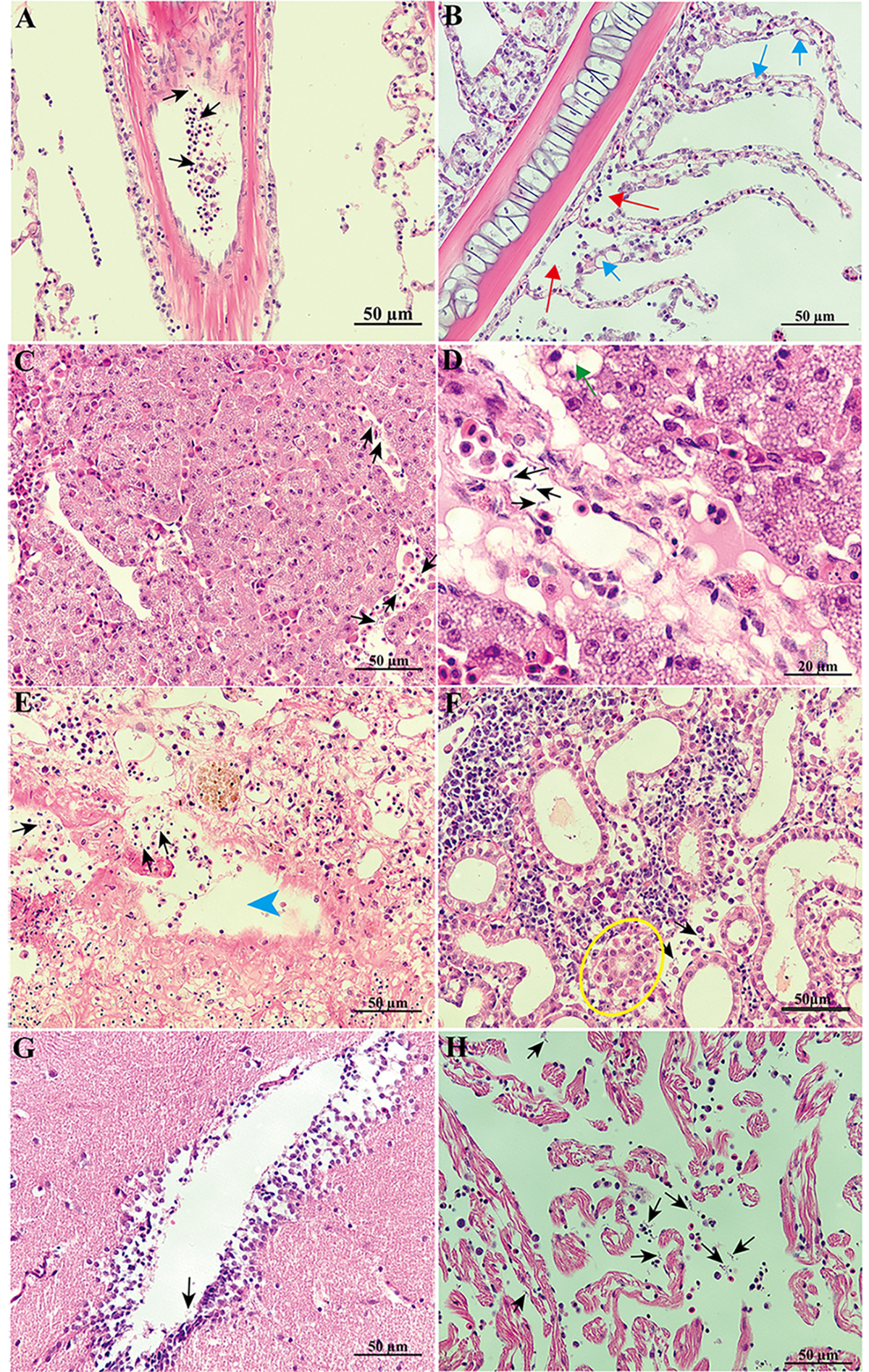
Histology of L. crocea infected with trypanosomes. A: trypanosomes (black arrow) were observed in vessels of the gill. B: hyperplasia (blue arrow) and gill epithelial detachment (red arrow) were evident in secondary lamellae. C: a massive amount of trypanosomes (black arrow) were inside the vessels of the liver. D: vacuolated hepatocytes (green arrow) were observed in the liver. E: trypanosomes (black arrow) were inside the spleen and atrophy of the white pulp was evident (blue arrowhead) in the areas near the location where the trypanosomes were located. F: the tubular epithelial cells in the kidney were detached (yellow cycle). Parasites were found in the brain (G) and heart (H). All scale bars = 50 μm (except in D, scale bar = 20 μm).
Current usage metrics show cumulative count of Article Views (full-text article views including HTML views, PDF and ePub downloads, according to the available data) and Abstracts Views on Vision4Press platform.
Data correspond to usage on the plateform after 2015. The current usage metrics is available 48-96 hours after online publication and is updated daily on week days.
Initial download of the metrics may take a while.