Figure 6
Download original image
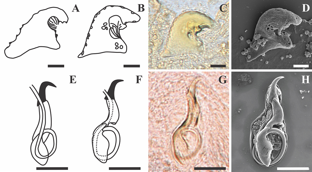
Line drawings, light and scanning electron micrographs of the male copulatory organ and vagina of Dactylogyrus teresae Mashego, 1983 from this study compared with line drawings from the original description. (A & E) – line drawings of vagina and MCO redrawn from Mashego (1983); (B & F) – line drawings of vagina and MCO from present study; (C & G) – light micrograph of vagina and MCO from present study; (D & H) – scanning electron micrograph of vagina and MCO (ventral view) from previously mounted GAP specimens in present study (scale value for all drawings and micrographs of vagina 5 μm and MCO 10 μm).
Current usage metrics show cumulative count of Article Views (full-text article views including HTML views, PDF and ePub downloads, according to the available data) and Abstracts Views on Vision4Press platform.
Data correspond to usage on the plateform after 2015. The current usage metrics is available 48-96 hours after online publication and is updated daily on week days.
Initial download of the metrics may take a while.