Figure 5

Download original image
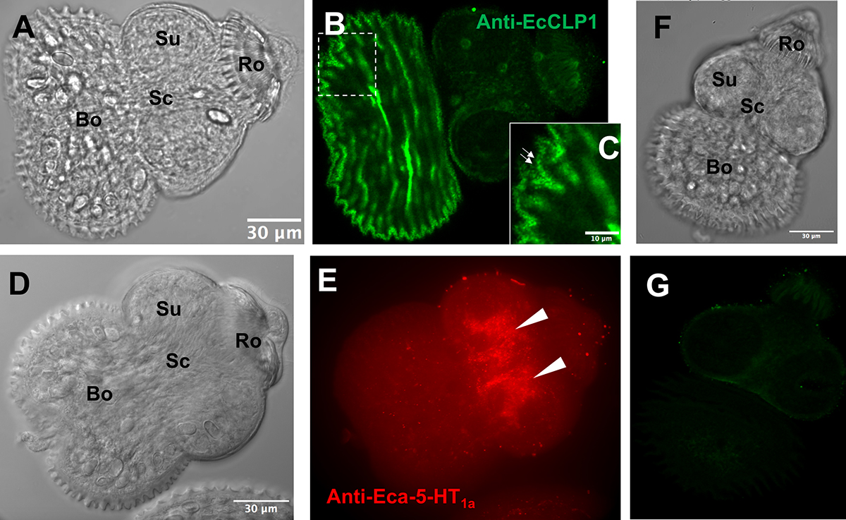
Immunolocalization of EcCLP1 in protoscoleces of Echinococcus canadensis. Protoscoleces were probed with anti-EcCLP1 (green) and anti-Eca-5-HT1a (red) serum and visualized by confocal microscopy. (A) Phase contrast view of the protoscolex shown in (B). (B) Fluorescent image of the protoscolex labeled with anti-EcCLP1 hyperimmune serum. Intense signals like dots were found superficially in the body region. (C) Magnification of the panel (B) showing a dotted pattern of staining on the surface of the tegument. (D) Phase contrast view of the protoscolex shown in (E). (E) Fluorescent image of the protoscolex labeled with anti-Eca-5-HT1a hyperimmune serum. (F) Phase contrast view of the protoscolex shown in (G). (G) Fluorescent image of the protoscolex labeled with the preimmune rabbit serum. The white arrows show the localization of Eca-5-HT1a in the cerebral ganglia in panel E or the localization of EcCLP1 in small dots in panel (C), suggesting a secretory release of the protein. Abbreviations, Bo: body region, Ro: rostellum, Sc: scolex region, Su: sucker.
Current usage metrics show cumulative count of Article Views (full-text article views including HTML views, PDF and ePub downloads, according to the available data) and Abstracts Views on Vision4Press platform.
Data correspond to usage on the plateform after 2015. The current usage metrics is available 48-96 hours after online publication and is updated daily on week days.
Initial download of the metrics may take a while.


