Figure 3

Download original image
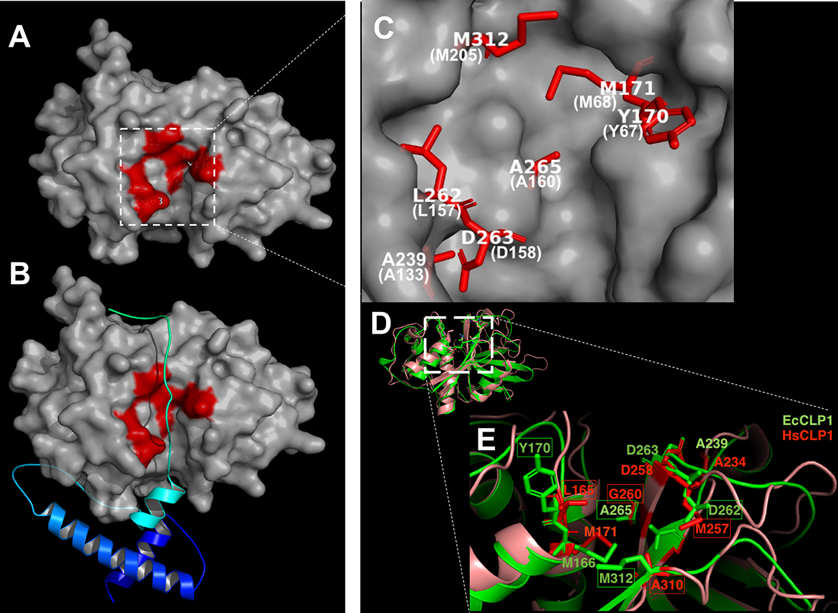
Predicted three-dimensional structures of proEcCLP1 and mature EcCLP1 and comparison with the human cathepsin. (A) Topological representation of the mature EcCLP1 with the residues forming the S2 subsite marked in red. (B) proEcCLP1 showing the propeptide in cyan and blue. (C) Magnified image of the model shown in (A) showing the residues forming the S2 subsite in white with papain numbering in parentheses. (D) Superposition of the ribbon representation of the crystal structure of human cathepsin (brown) [6] and the modeled EcCLP1 (green). (E) Magnified image of the active site of the cathepsins shown in (D). Important S2 residues are marked in green in parasite numbering and red in human cathepsin. Differences between both sequences are marked with boxes.
Current usage metrics show cumulative count of Article Views (full-text article views including HTML views, PDF and ePub downloads, according to the available data) and Abstracts Views on Vision4Press platform.
Data correspond to usage on the plateform after 2015. The current usage metrics is available 48-96 hours after online publication and is updated daily on week days.
Initial download of the metrics may take a while.


