Figure 2
Download original image
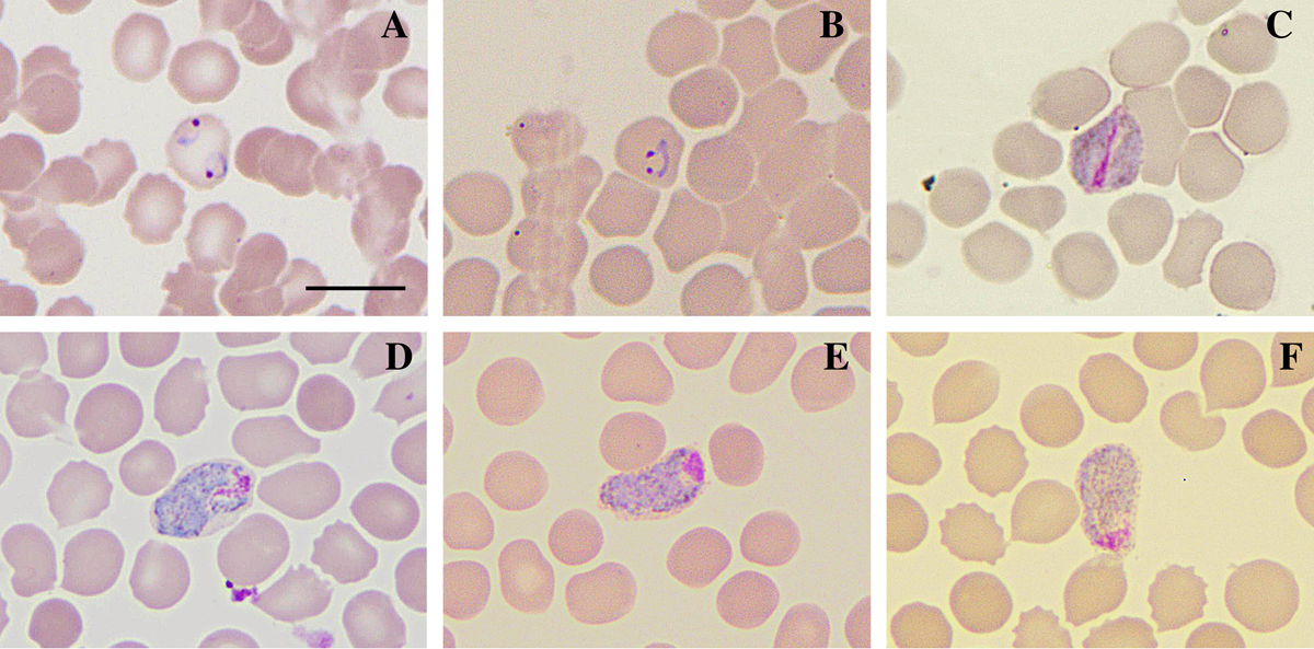
Atypical P. vivax morphologies found in the sample CAM-190. Slides were prepared by the blood distension technique, stained by the Giemsa method, and visualized at 100× magnification under an optical microscope. (A) multi-infected red blood cell with trophozoites; (B) binucleated trophozoite; (C) band-form gametocyte; (D–F) elongated gametocytes resembling a sausage shape. Bar = 10 μm.
Current usage metrics show cumulative count of Article Views (full-text article views including HTML views, PDF and ePub downloads, according to the available data) and Abstracts Views on Vision4Press platform.
Data correspond to usage on the plateform after 2015. The current usage metrics is available 48-96 hours after online publication and is updated daily on week days.
Initial download of the metrics may take a while.