Figure 4
Download original image
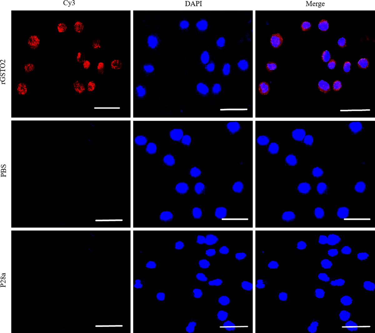
Localization of F. hepatica-derived rGSTO2 protein to the RAW264.7. macrophages surface. The macrophages were respectively pre-treated with rGSTO2, PBS or P28a protein, and the cells were then incubated with mouse anti-rGSTO2 IgG, naïve mouse serum or mouse anti-P28a IgG as primary antibody, followed by Cy3-conjugated goat anti-mouse IgG as the secondary antibody. The red fluorescence on the surface of cells indicated that the target protein (rGSTO2 protein) is stained (Cy3) and the cell nuclei was stained by DAPI (blue), while merge represents the overlap of the red and blue fluorescence channels. No fluorescence was observed in PBS or P28a control groups. Scale-bars: 10 μm.
Current usage metrics show cumulative count of Article Views (full-text article views including HTML views, PDF and ePub downloads, according to the available data) and Abstracts Views on Vision4Press platform.
Data correspond to usage on the plateform after 2015. The current usage metrics is available 48-96 hours after online publication and is updated daily on week days.
Initial download of the metrics may take a while.