Figure 1

Download original image
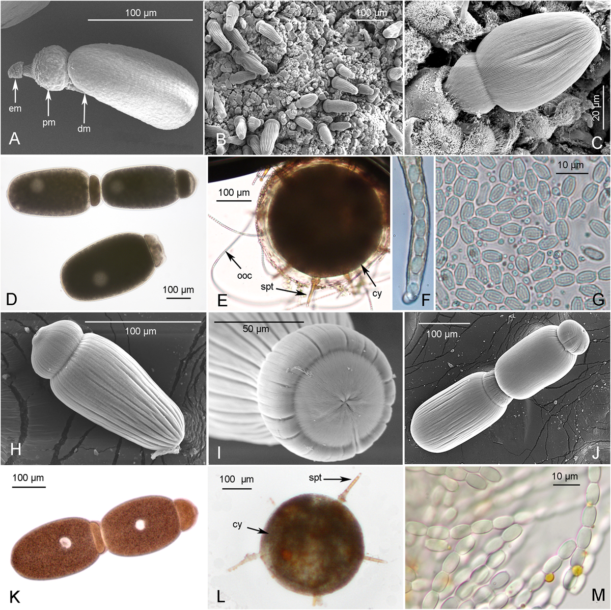
Scanning Electron Microscopy (A–C, H–J) and photonic imaging (D–G, K–M) of gregarines infecting S. gregaria (A–G) and L. migratoria (H–M). S. gregaria gregarines: A, young trophozoite (epimerite (em) protomerite (pm) and deutomerite (dm)), (South Africa); B, intestinal tract infected by numerous gregarines (Morocco); C, gregarine encased in an intestinal host cell, enlargement of B (Morocco); D. Solitary gamont and syzygy (Belgium); E. Gametocyst form (cy) with developed sporoducts (spt) releasing oocyst chains (ooc); F, zoom on sporoduct extremity showing enclosed oocysts; G. released oocysts. L. migratoria gregarines: H, solitary gamont detached from intestinal host cell; I. zoom on gamont protomerite; J–K, gamonts associated in syzygies; L, Gametocyst form (cy) with developed sporoducts (spt); M. released oocysts. Scales are given for each figure.
Current usage metrics show cumulative count of Article Views (full-text article views including HTML views, PDF and ePub downloads, according to the available data) and Abstracts Views on Vision4Press platform.
Data correspond to usage on the plateform after 2015. The current usage metrics is available 48-96 hours after online publication and is updated daily on week days.
Initial download of the metrics may take a while.


