Figure 2
Download original image
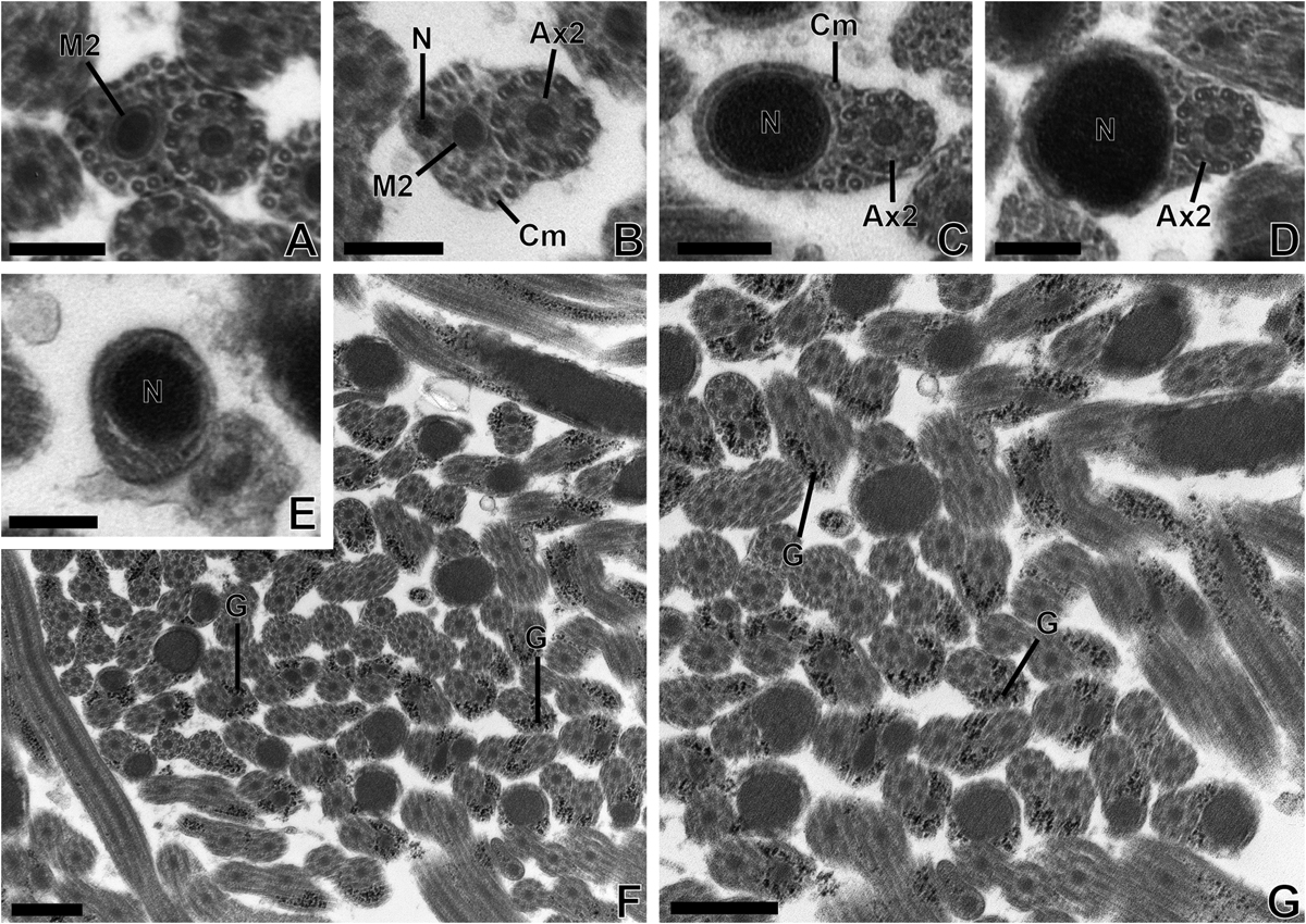
(A–E) Transmission electron micrographs of region IV (posterior region) of the mature spermatozoon of Tergestia clonacantha. Scale bars = 0.2 μm. Cross-sections in region IV of mature spermatozoon of Tergestia clonacantha showing the appearance of the second mitochondrion (M2) (A), then the nucleus (N) (B). (C) Cross-sections showing the disappearance of the first mitochondrion, a progressive disappearance of microtubules (C–D) and axoneme 2 (E). Then, the posterior extremity of the spermatozoon exhibiting only the nucleus (E). (F–G) Transmission electron micrographs showing many sections of the mature spermatozoon of Tergestia clonacantha with granules of glycogen (G) highlighted by the Thiéry method. Scale bars = 0.5 μm.
Current usage metrics show cumulative count of Article Views (full-text article views including HTML views, PDF and ePub downloads, according to the available data) and Abstracts Views on Vision4Press platform.
Data correspond to usage on the plateform after 2015. The current usage metrics is available 48-96 hours after online publication and is updated daily on week days.
Initial download of the metrics may take a while.