Figure 2A and B
Download original image
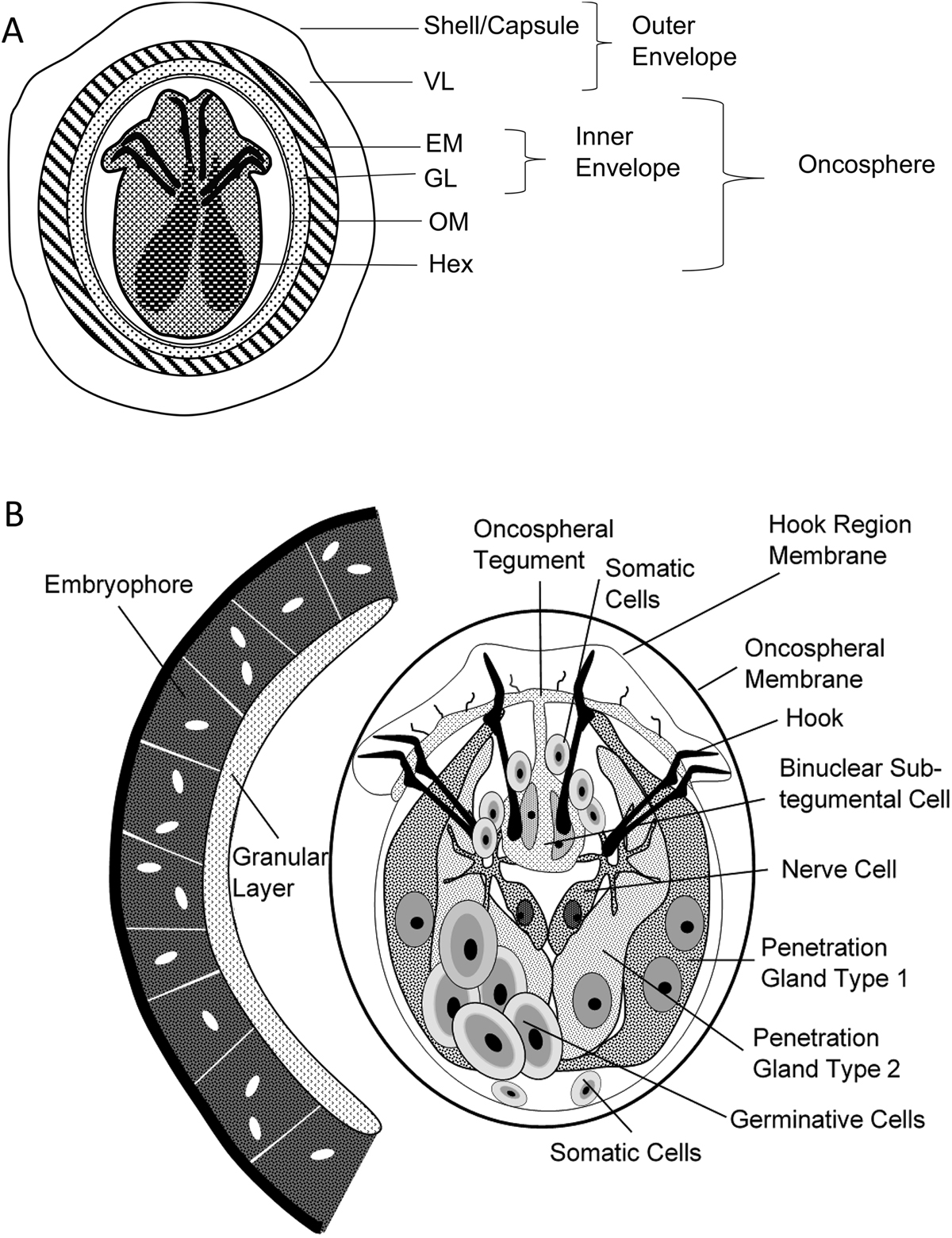
General description of the egg and oncosphere of Echinococcus spp, according to Jabbar et al., 2010 [27]. (A) Schematic diagram of an oncosphere illustrating the structure and bilateral symmetry in the pattern of hooks and cellular organization of the hexacanth embryo. VL: vitelline layer; EM: embryophore; GL: granular layer; OM: oncospheral membrane; Hex: hexacanth embryo. (B) Cellular organization of the oncosphere. Oncospheres are approximately 25 × 30 μm.
Current usage metrics show cumulative count of Article Views (full-text article views including HTML views, PDF and ePub downloads, according to the available data) and Abstracts Views on Vision4Press platform.
Data correspond to usage on the plateform after 2015. The current usage metrics is available 48-96 hours after online publication and is updated daily on week days.
Initial download of the metrics may take a while.