Figure 4
Download original image
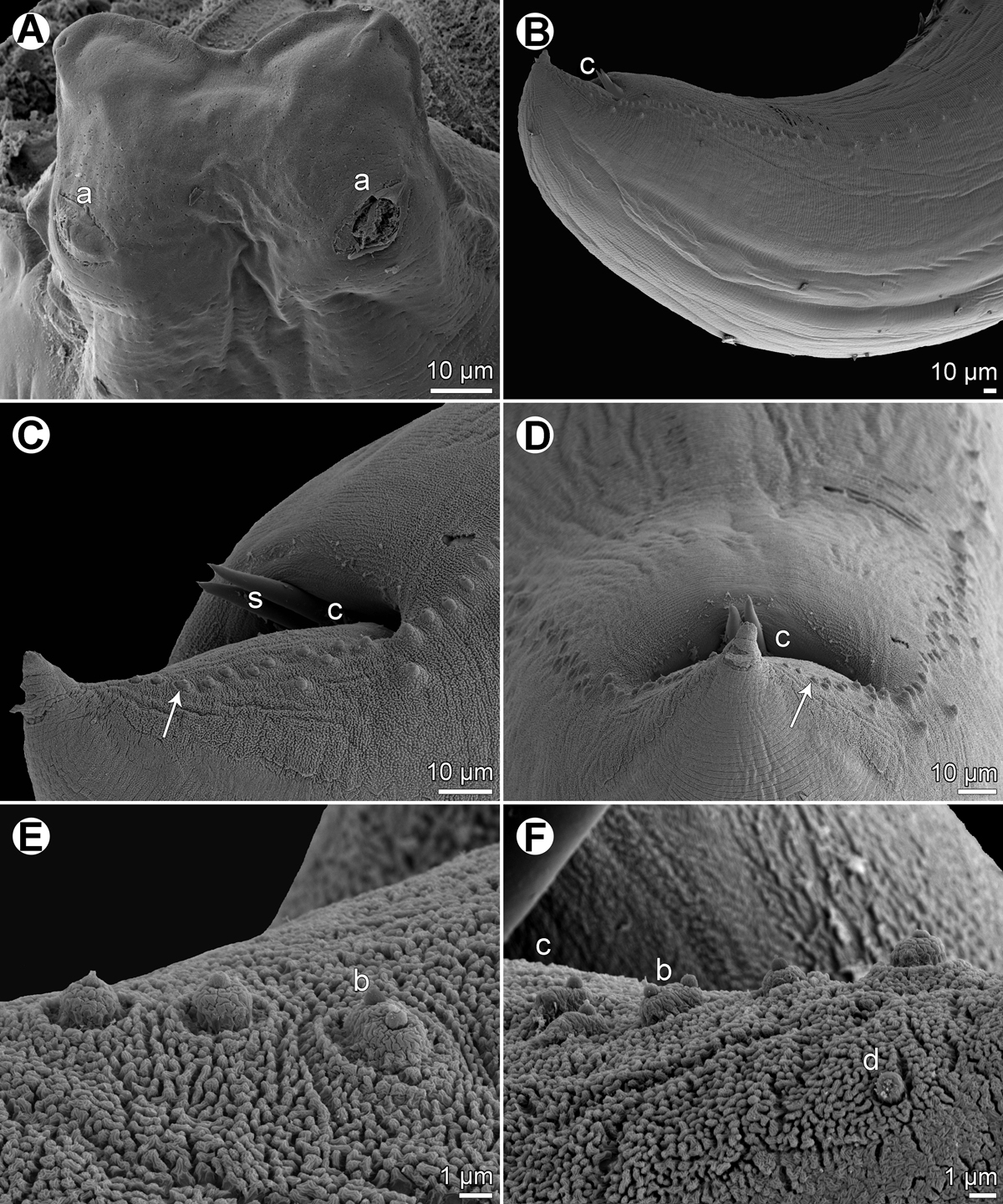
Raphidascaris (Ichthyascaris) fasciati n. sp., scanning electron micrographs of male. (A) Dorsal lip; (B) posterior end of male, lateral view; (C) tail, sublateral view (arrow indicates postanal double papilla); (D) region of cloaca and tail, dorsoventral view (arrow indicates postanal double papilla); (E) postanal papillae of three posteriormost pairs; (F) region of postanal papillae of four posteriormost pairs, lateral view. (a) Labial double papilla; (b) postanal double papilla; (c) cloacal aperture; (d) phasmid; (s) spicules.
Current usage metrics show cumulative count of Article Views (full-text article views including HTML views, PDF and ePub downloads, according to the available data) and Abstracts Views on Vision4Press platform.
Data correspond to usage on the plateform after 2015. The current usage metrics is available 48-96 hours after online publication and is updated daily on week days.
Initial download of the metrics may take a while.