Figure 8
Download original image
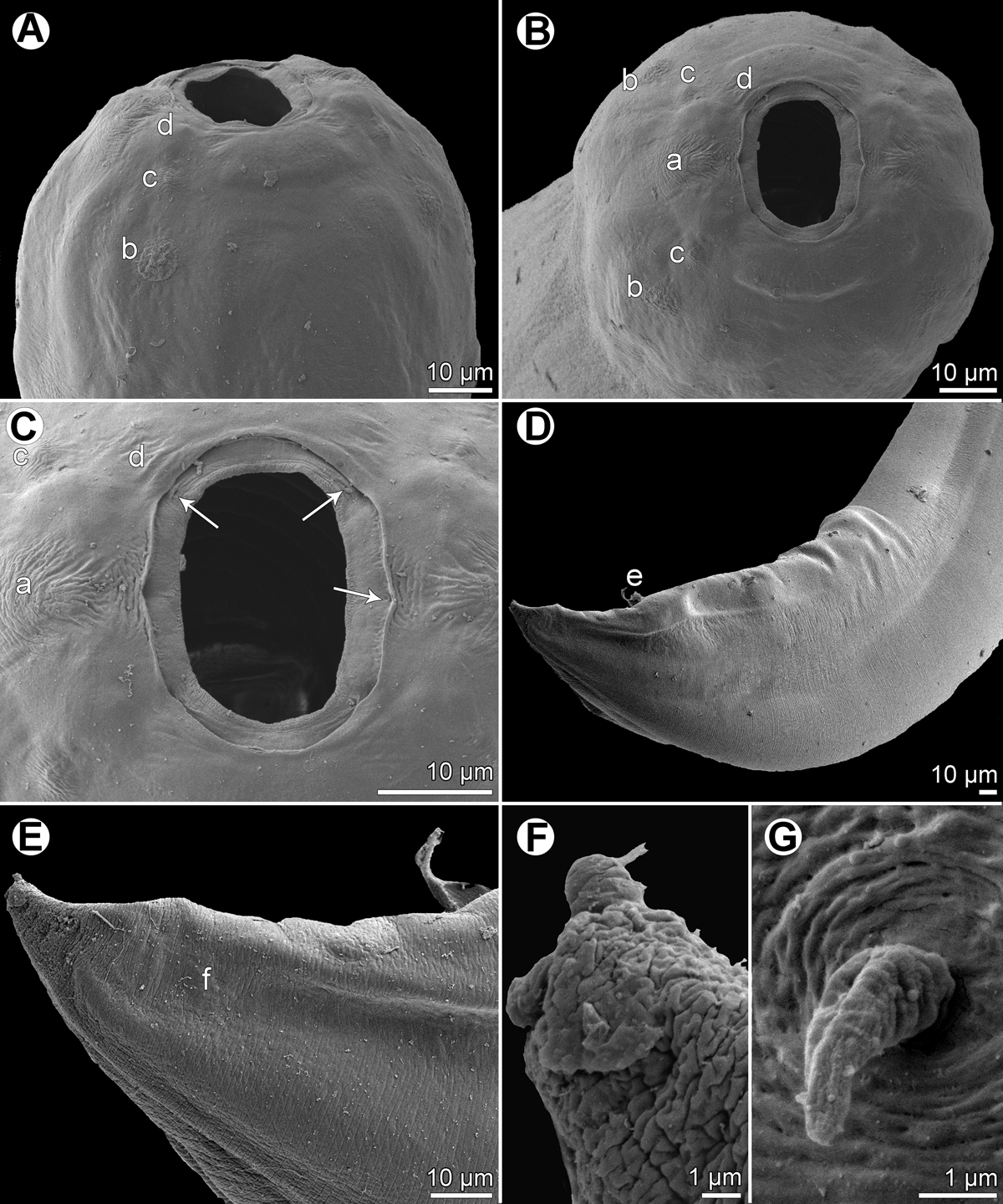
Procamallanus (Spirocamallanus) synodi n. sp., scanning electron micrographs. (A, B) Cephalic end, subdorsoventral and apical views, respectively; (C) region of oral aperture, apical view (arrows indicate circumoral pores); (D) posterior end of male, lateral view; (E) tail of male, lateral view; (F) tail tip of female, lateral view; (G) deirid. (a) amphid; (b) cephalic papilla of external circle; (c) cephalic papilla of middle circle; (d) cephalic papilla of internal circle; (e) cloaca; (f) phasmid.
Current usage metrics show cumulative count of Article Views (full-text article views including HTML views, PDF and ePub downloads, according to the available data) and Abstracts Views on Vision4Press platform.
Data correspond to usage on the plateform after 2015. The current usage metrics is available 48-96 hours after online publication and is updated daily on week days.
Initial download of the metrics may take a while.