Figure 2
Download original image
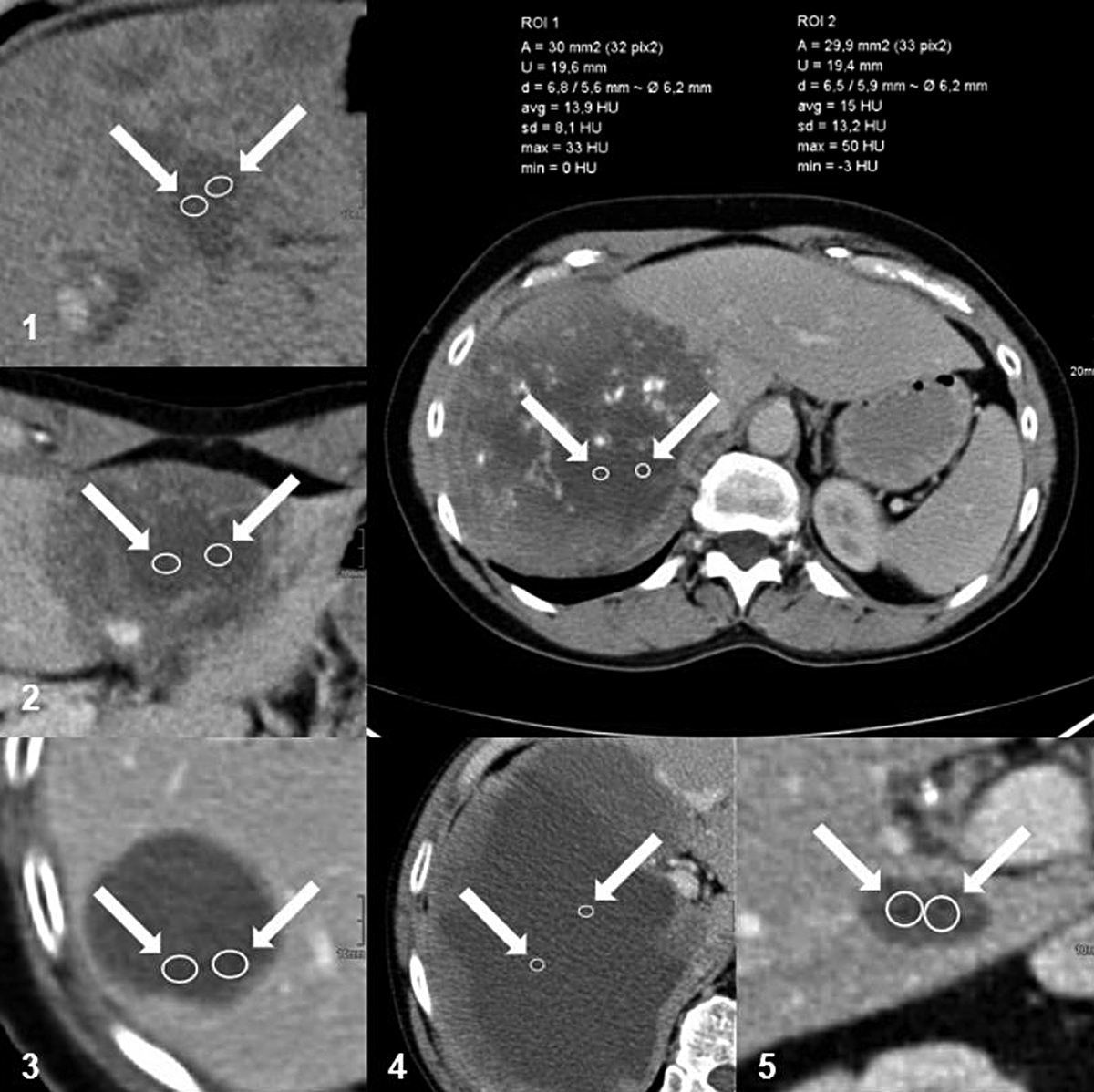
Example of density measurement in the cystoid portions of AE lesions on CT. Left and below: in each case, two enlarged ROIs within the cystoid portions of AE lesions of different primary morphological types: (1) Type I “diffuse infiltrating with cystoid portion”; (2) Type II “primarily circumscribed tumour-like with cystoid portion”; (3) Type IIIa “primarily cystoid intermediate without solid portion at the edge”; (4) Type IIIb “primarily cystoid widespread with solid portion at the edge”; (5) Type IV “small cystoid/metastatic”. Above right: two ROIs with surface data and HU values (max, min, mean and standard deviation) within the cystoid portion of an AE lesion involving both lobes of the liver. (AE = alveolar echinococcosis; CT = computed tomography; HU = Hounsfield unit; ROI = region of interest).
Current usage metrics show cumulative count of Article Views (full-text article views including HTML views, PDF and ePub downloads, according to the available data) and Abstracts Views on Vision4Press platform.
Data correspond to usage on the plateform after 2015. The current usage metrics is available 48-96 hours after online publication and is updated daily on week days.
Initial download of the metrics may take a while.